Category: Neurology
Keywords: thrombolytics, acute ischemic stroke, stroke, hemorrhage, dental procedures, minor surgery (PubMed Search)
Posted: 8/3/2011 by Aisha Liferidge, MD
Click here to contact Aisha Liferidge, MD
Category: Critical Care
Keywords: trauma, resuscitaiton, pregnancy, IVC, supine hypoventilation, edema, intubation, RSI, desaturaiton (PubMed Search)
Posted: 8/2/2011 by Haney Mallemat, MD
Click here to contact Haney Mallemat, MD
Pregnancy causes many physiologic changes, which may be challenging during trauma resuscitations. A few pearls on the ABC’s:
Airway
Breathing
Circulation
Chesnutt, A. Physiology of normal pregnancy. Crit Care Clinics 2004 Oct;20(4):609-15.
Follow me on Twitter (@criticalcarenow) or Google+ (+haney mallemat)
Category: Medical Education
Keywords: Interns, Emergency Medicine (PubMed Search)
Posted: 8/1/2011 by Rob Rogers, MD
(Updated: 2/8/2026)
Click here to contact Rob Rogers, MD
In a 1991 article published by Wrenn and Slovis, the ten commandments of emergency medicine are discussed. This is an awesome article and a must read for everyone in EM.
Michelle Lin recently mentioned these on her blog, Academic Life in Emergency Medicine....
The Ten Commandments of Emergency Medicine:
1. Secure the ABC's
2. Consider or give naloxone, glucose, and thiamine
3. Get a pregnancy test
4. Assume the worst
5. Do not send unstable patients to radiology
6. Look for common red flags
7. Trust no one, believe nothing (not even yourself)
8. Learn from your mistakes
9. Do unto others as you would do to your family (and that includes coworkers)
10. When in doubt, always err on the side of the patient
These are great teaching pearls for new interns and for the rest of us as well....
Wrenn, Slovis
Michelle Lin's blog, Academic Life in Emergency Medicine
Category: Misc
Keywords: Wound, Repair (PubMed Search)
Posted: 7/30/2011 by Michael Bond, MD
Click here to contact Michael Bond, MD
Wound Repair
A pearl last year addressed the irrigation of wound and the fact that the type of fluid (sterile versus tap water) does not affect infection rates but rather the volume of irrigation is most important.
Sterile versus unsterile gloves have also been studied, and it turns out that clean unsterile gloves have the same rate of infection as sterile gloves but come with a substantial cost savings.
When caring for a contaminated wound it is most important to remove any gross contamination, and then irrigate the wound as much as possible. A 20 mL syringe with an 18G angio-catheter provides the proper pressure to remove debris without causing tissue damage. The wound can then be closed wearing the gloves that are most comfortable or accessible to you.
Finally, from a medicolegal standpoint it is always best to inform the patient that you have tried to remove all of the contamination but there is still a chance that the wound can get infected.
Category: Pediatrics
Posted: 7/29/2011 by Mimi Lu, MD
Click here to contact Mimi Lu, MD
Acute Poststreptococcal Glomerulonephritis (APSGN) is a sequela of group A beta-hemolytic streptococci (GAS) infection of the skin or pharynx with nephrogenic strains of GAS. Damage to the kidneys is due to deposition of antigen-antibody complexes in the glomeruli.
Presentation:
Testing:
Treatment:
Prognosis (favorable):
Reference:
Kit, Brian. Assess the volume status and electrolytes in children with poststreptococcal glomerulonephritis. Avoiding Common Pediatric Errors. 2008. p356-57.
Category: Toxicology
Keywords: fluroquinolone, tendon rupture (PubMed Search)
Posted: 7/28/2011 by Bryan Hayes, PharmD
Click here to contact Bryan Hayes, PharmD
The incidence of tendon rupture related to fluoroquinolone use is reported to be in the range of 1 in 6000.
The risk of tendon rupture associated with FQ use is increased in those older than 60 years of age, those taking steroids, and in patients who have received heart, renal, or pulmonary transplants.
There is no evidence that tendon rupture is more likely for patients taking levofloxacin compared to other FQs.
The Medical Letter 2011;53(1368):55-56.
Category: Neurology
Keywords: central pontine myelinolysis, hypernatremia (PubMed Search)
Posted: 7/27/2011 by Aisha Liferidge, MD
(Updated: 2/8/2026)
Click here to contact Aisha Liferidge, MD
Category: Critical Care
Posted: 7/26/2011 by Mike Winters, MBA, MD
Click here to contact Mike Winters, MBA, MD
Blood Pressure in the Critically Ill Obese Patient
King DR, Velmahos GC. Difficulties in managing the surgical patient who is morbidly obese. Crit Care Med 2010; 38(S):S478-82.
Category: Visual Diagnosis
Posted: 7/25/2011 by Haney Mallemat, MD
(Updated: 8/28/2014)
Click here to contact Haney Mallemat, MD
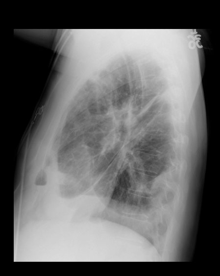
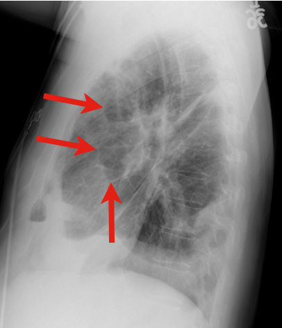
34 y.o. male with history of IVDA (intravenous drug abuse) complains of fever, chills and cough. Diagnosis?

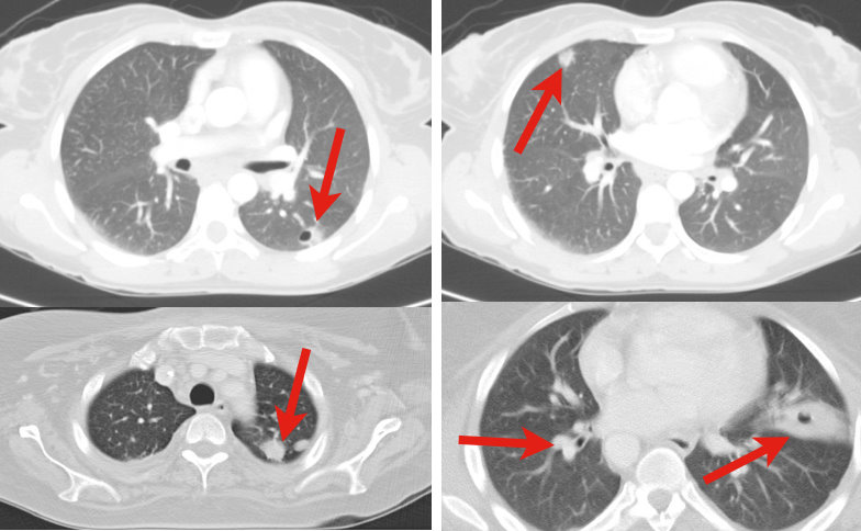
Answer: Lung Abscess (from septic pulmonary emboli)

Lung Abscess
Necrosis of lung parenchyma with pus and debris-filled cavities
Caused by direct injury (e.g., aspiration pneumonia) or secondary causes (e.g., tricuspid endocarditis, bacteremia, etc.)
Suspect with:
Loss of airway reflexes (e.g., CVA, seizures, alcohol / narcotic abuse, etc)
Poor dentition
Immunosuppression
IVDA
Gram positives, negatives and anaerobic bacteria have all been implicated.
CXR may suggest diagnosis, but CT scan better identifies abscess, necrotic tissue, empyema, or other pathology (see image below).
After drawing blood cultures, broad-spectrum antibiotics should be started and narrowed once culture data is available; address underlying cause (e.g., valve replacement for endocarditis).
Prognosis is generally good with normal immune function and antibiotics, but mortality sharply increases with immunocompromise and treatment delay.

Mansharamani N, et al. Lung abscess in adults: clinical comparison of immunocompromised to non-immunocompromised patients. Respiratory Medicine. Mar 2002;96(3):178-85
Follow me on Twitter (@criticalcarenow) or Google+ (+haney mallemat)
Category: Cardiology
Keywords: myocardial infarction, right ventricle, right ventricular (PubMed Search)
Posted: 7/24/2011 by Amal Mattu, MD
Click here to contact Amal Mattu, MD
Clues to RV infarction:
1. This almost always occurs in the presence of a concurrent inferior MI.
2. Clinical findings may include the triad of hypotension, JVD, and clear lungs.
3. ECG clues: in the presence of inferior lead ischemia or injury pattern, look for:
a. Combination of ST depression in lead V2 + ST elevation in lead V1; OR
b. Combination of ST depression in lead V2 + isoelectric ST segments in leads V1 and V3; OR
c. ST elevation in lead III markedly greater than the ST elevation in lead II; OR
d. ST elevation in right-sided leads (requires you to obtain right-sided leads)
Why is this diagnosis important?
1. It suggests a larger infarction and worse prognosis, so BE AGGRESSIVE in management.
2. Be very cautious with preload-reducing medications (e.g. nitrates) in the acute management of these patients, as they may induce significant reductions in blood pressure and extension of the infarction. Be aggressive with IVF, while maintaining close attention to the lung sounds.
Category: Orthopedics
Keywords: Osteomyelitis, hyperbaric oxygen (PubMed Search)
Posted: 7/23/2011 by Brian Corwell, MD
Click here to contact Brian Corwell, MD
Refractory Osteomyelitis is defined as a chronic osteomyelitis that persists or recurs after appropriate interventions have been performed or where acute osteomyelitis has not responded to surgery and antibiotics.
Case series, animal data and non-randomized prospective trials suggest that the addition of Hyperbaric Oxygen therapy to routine surgical and antibiotic management of previously refractory osteomyelitis is safe and improves the rate of infection resolution.
In patients with osteomyelitis involving spine, skull, sternum, HBOT is recommended prior to surgical intervention.
Typically patients require 20-40 daily dives for sustained therapeutic benefit.
How does HBOT work in osteomyelitis?
1. Restoration of normal to elevated O2 level in infected bone.
2. Leukocyte mediated killing of aerobic bacteria is restored when low O2 tension intrinsic to osteomyolitic bone is restored to physiologic or supra-physiologic levels.
3. HBOT is noted to exert direct suppressive effects on anaerobic infections.
4. HBOT augment the transport of certain abx (aminoglycosides and cephalosporins) across bacterial cell wall.
5. Enhance osteogenesis
6. Enhance angiogenesis
thank you to Dr. Sethuraman for this pearl
“Refractory Osteomyelitis” written by Hart, Brett, MD In: Hyperbaric Oxygen Therapy Indications Editor: Gesell, Laurie, MD FACEP
Category: Pediatrics
Posted: 7/22/2011 by Mimi Lu, MD
Click here to contact Mimi Lu, MD
You're called to bedside to evaluate a "lethargic" infant. You wisely ask for a POCT glucose which returns at 35. How much dextrose do you give (since you know it's not just "an amp" of D50?
Here's a simple mnemonic:
Rule of 50-100 = multiply type of dextrose solution by ____ factor (ml/kg) to total 50-100
D10 (neonate) x 5-10 ml/kg = 50-100
D25 (infant) x 2-4 ml/kg = 50-100
D50 (child/adolescent) x 1-2 ml/kg = 50-100
Category: Toxicology
Keywords: mephedrone (PubMed Search)
Posted: 7/21/2011 by Fermin Barrueto
(Updated: 2/8/2026)
Click here to contact Fermin Barrueto
There are increasing reports of bath salts which are crushed and then either injected, insufflated or taken orally. The actual substance has been found to be mephedrone as well as MDPV.(1) Both are amphetamine derivatives and the psychosis seen can appear like schizophrenia to the point that some of these patients have been admitted to the psychiatric wards. (2)(3) For those who have seen methamphetamine patients "tweaking" - where they use the drug for several days in a row without sleep - the presentation is quite similiar.
Synthetic drugs continue to present legal and regulatory problems since the compound is a "designer" synthesized drug that may not be on the DEA Schedule list. The product is labeled "Not for human consumption". Head shops and the internet remain primary sources of the drug. Bath salts present a serious and dangerous public health risk.
1) Centers for Disease Control and Prevention (CDC). Emergency department visits after use of a drug sold as "bath salts"--Michigan, November 13, 2010-March 31,2011. MMWR Morb Mortal Wkly Rep. 2011 May 20;60(19):624-7.
2) Antonowicz JL, Metzger AK, Ramanujam SL. Paranoid psychosis induced by consumption of methylenedioxypyrovalerone: two cases. Gen Hosp Psychiatry. 2011 May 25.
3) Penders TM, Gestring R. Hallucinatory delirium following use of MDPV: "Bath Salts". Gen Hosp Psychiatry. 2011 Jul 13. [Epub ahead of print] PubMed PMID: 21762997.
Category: Neurology
Keywords: PRES, posterior reversible encephalopathy syndrome (PubMed Search)
Posted: 7/20/2011 by Aisha Liferidge, MD
Click here to contact Aisha Liferidge, MD
Category: Critical Care
Keywords: heat stroke, critical care, acute kidney injury, seizures, neurological (PubMed Search)
Posted: 7/19/2011 by Haney Mallemat, MD
Click here to contact Haney Mallemat, MD
Heat stroke is hyperthermia (>41.6 Celsius / 106 Fahrenheit) plus neurologic findings (e.g., altered mental status, seizures, coma, etc.); it also causes systemic inflammation response syndrome (i.e., cytokine release), coagulation disorders (e.g., thrombosis in end organs) and tissue abnormalities (e.g., acute kidney injury and rhabdomyolysis)
Two classifications exist:
Treatment includes:
Despite the most aggressive therapy, up to 30% survivors may have permanent neurologic or multi-organ system dysfunction months to years after recovery
Leon, L. Heat stroke: role of the systemic inflammatory response. Journal of Applied Physiology 2010 Dec;109(6):1980-8
http://emedicine.medscape.com/article/166320-overview
Follow me on Twitter: @criticalcarenow or Google +: @haney mallemat
Category: Cardiology
Keywords: phenylephrine (PubMed Search)
Posted: 7/17/2011 by Amal Mattu, MD
(Updated: 2/8/2026)
Click here to contact Amal Mattu, MD
With recent national shortages of norepinephrine, our typical go-to drug in sepsis, it's become important for us all to familiarize ourselves with alternative pressors in this setting. Phenylephrine is a commonly chosen alternative.
Phenylephrine is a potent alpha-agonist associated with peripheral vasoconstriction. It has no beta effects so it is not associated with tachydysrhythmias. On the other hand, it is associated with reflex bradycardia which can be treated or prevented with atropine (although there are no specific recommendations to routinely administer atropine prophylactically). Phenylephrine may take 10 minutes to demonstrate an effect, and its duration is approximately 15 minutes. It should be used cautiously in patients with underlying cardiac disease because of the vasoconstrictive effect, and it should be avoided in patients with narrow-angle closure glaucoma.
Extravasation can cause tissue necrosis and should be treated with phentolamine.
Category: Infectious Disease
Keywords: C. Diff Colitis (PubMed Search)
Posted: 7/16/2011 by Michael Bond, MD
(Updated: 2/8/2026)
Click here to contact Michael Bond, MD
C. Diff Colitis
The general treatment recommendations for C. Diff Colitis are to place the patient on PO metronidazole and if they fail this treatment PO vancomycin (125 mg 4x day). Vancomycin is generally reserved for resistant cases due to the fear that it could induce Vancomycin resistant enterococcus.
For severally ill patients it is recommended that you prescribe IV metronidazole and PO vancomycin when they are not actively vomiting. Remember there is no role for IV vancomycin as it does not get into the bowel lumen to eradicate the infection.
There is some great news though, the FDA recently approved a new drug, a macrolide antibiotic fidaxomicin (Dificid), for the treatment of C. Diff Colitis. Fidaxomicin was found to be as effective as vancomycin in preventing recurrence 3 weeks after treatment. Currently it is recommended that fidaxomicin be reserved for cases where patients are having recurrences after 3 weeks of vancomycin treatment.
The FDA news release can be found at http://www.fda.gov/NewsEvents/Newsroom/PressAnnouncements/ucm257024.htm
Category: Pediatrics
Keywords: Enterovirus, infant, CSF (PubMed Search)
Posted: 7/15/2011 by Mimi Lu, MD
(Updated: 7/22/2011)
Click here to contact Mimi Lu, MD
Now that summer is in full swing, the question is: Should the evaluation of the febrile young infant change during the summer and fall months? And can that affect length of hospitalization and antibiotic use?
Two retrospective cohort studies from the Children’s Hospital of Philadelphia (CHOP) suggest yes! The addition of enterovirus polymerase chain reaction (PCR) testing to cerebrospinal fluid (CSF) may improve the care of infants with fever during enterovirus season (early June through late October).
Of note, at CHOP: 1) infants 56 days or younger routinely undergo lumbar puncture during evaluation for fever. 2) Most CSF enterovirus PCR test results (90%) were available within 36 hours; 95% of results were available within 48 hours.
In the King study, having positive enterovirus PCR CSF results decreased the length of hospitalization and the duration of antibiotic use for young infants less than 90 days, supporting the routine use of this test during periods of peak enterovirus season. In multivariate
analysis, a positive CSF enterovirus PCR result was associated with a 1.54-day decrease in the length of stay and a 33.7% shorter duration of antibiotic use.
Bottom line: Consider adding enterovirus PCR testing to CSF obtained during the evaluation of febrile young infants during enterovirus season, as this may reduce length of hospitalization and duration of antibiotic use. The effects, however, may be limited at institutions with slower lab turnaround times.
References:
1) King RL, Lorch SA, Cohen DM, Hodinka RL, Cohn KA, Shah SS. Routine cerebrospinal fluid enterovirus polymerase chain reaction testing reduces hospitalization and antibiotic use for infants 90 days or younger. Pediatrics. 2007 Sep;120(3):489-96. http://pediatrics.aappublications.org/content/120/3/489.full.pdf
2) Dewan M, Zorc JJ, Hodinka RL, Shah SS. Cerebrospinal fluid enterovirus testing in infants 56 days or younger. Arch Pediatr Adolesc Med. 2010 Sep;164(9):824-30.
Category: Toxicology
Keywords: levamisole, cocaine, vasculitis, agranulocytosis, heroin (PubMed Search)
Posted: 6/23/2011 by Bryan Hayes, PharmD
(Updated: 7/14/2011)
Click here to contact Bryan Hayes, PharmD
Levamisole is an antihelminthic agent used in humans to treat certain parasitic infections and cancers. It is more commonly used for veterinary purposes. It has recently seen increasing use as a cutting agent for cocaine and heroin, found in up to 70% of cocaine sample seized by the DEA. It adds bulk and weight to powdered cocaine and is even theorized to increase the stimulant effects.
Toxicity of levamisole includes agranulocytosis and vasculitis (see attached document for recent image from NEJM).
Trivia: Levamisole was found in DJ AM and Andrew Koppel (Ted Koppel’s son), who both died of drug overdoses.
Category: Neurology
Keywords: ms, multiple sclerosis, plasmapharesis (PubMed Search)
Posted: 7/13/2011 by Aisha Liferidge, MD
(Updated: 2/8/2026)
Click here to contact Aisha Liferidge, MD
(1) immunomodulatory therapy for the underlying immune disorder, often with high dose
intravenous (IV) steroids which speeds recovery, and
(2) management of symptoms through supportive measures and amelioration of risk factors
associated with precipitating acute exacerbations such as infection through aggressive use
of antibiotics. Treatment of fever with antipyretics also key as even small increases in
temperature can significantly affect conduction through partially demyelinated fibers.
