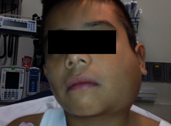
Category: Visual Diagnosis
Posted: 11/27/2011 by Haney Mallemat, MD
(Updated: 11/28/2011)
Click here to contact Haney Mallemat, MD
9 year-old boy with sudden onset of unilateral facial swelling. What’s the diagnosis?

Answer: Acute Parotitis
Bonus Trivia: U.S. President Garfield died from parotitis after becoming dehydrated following abdominal surgery
Shelly J. McQuone MD, Acute Viral and Bacterial Infections of the Salivary Glands, Otolaryngologic Clinics of North America, Volume 32, Issue 5 (October 1999)
Follow me on Twitter (@criticalcarenow) or Google+ (+haney mallemat)
Category: Cardiology
Keywords: endocarditis (PubMed Search)
Posted: 11/28/2011 by Amal Mattu, MD
(Updated: 2/9/2026)
Click here to contact Amal Mattu, MD
Right heart endocarditis is much more common in patients that are injection drug users. Fortunately for them, they have a lower mortality than patients with left heart endocarditis because they have a lower rate of developing heart failure. This is a reminder that the most common cause of death from endocarditis is heart failure.
Category: Orthopedics
Keywords: Weber, ankle fracture, fibula (PubMed Search)
Posted: 11/26/2011 by Brian Corwell, MD
(Updated: 2/9/2026)
Click here to contact Brian Corwell, MD
The Weber classification system
A commonly used, simple, easily remembered system used to describe ankle fractures. The system focuses on the integrity of the syndesmosis.
http://www.accessemergencymedicine.com/loadBinary.aspx?fileName=simo_c017f013t.gif
- TYPE A: fibula fracture below the ankle joint/syndesmosis (which is intact). Deltoid ligament intact. Medial malleolus can be fractured. Usually treated with closed reduction.
http://www.gentili.net/image1.asp?ID=-241442344&imgid=AnkleWeberAAP600.jpg&Fx=Weber+A+Fracture
- TYPE B: is a transsyndesmotic fracture with usually partial rupture of the syndesmosis (though may be intact). No gross widening to the tib/fib articulation.. Deltoid ligament intact. Medial malleolus often fractured. Variable stability. Any clinical or radiographic injury to the medial joint complex make this an unstable fracture
http://www.gentili.net/image.asp?ID=145&imgid=AnkleWeberBmortise600.jpg&Fx=Weber+B+Fracture
- TYPE C: Fibular fracture above the level of the syndesmosis with usually a total rupture of the syndesmosis (seen as widening of the distal tib/fin articulation), resulting in instability of the ankle mortise. Associated with medial malleolus fracture or deltoid ligament injury. Unstable.
http://www.gentili.net/image1.asp?ID=146&imgid=anklewebcapoblx2600.jpg&Fx=Weber+C+Fracture
Category: Pediatrics
Keywords: Kawasaki, vasculitis, fever, (PubMed Search)
Posted: 11/25/2011 by Mimi Lu, MD
Click here to contact Mimi Lu, MD
Classic Kawasaki is diagnosed by fever for greater than 5 days plus 4 out of 5 classic signs.
But what about an 8 month-old with 6 days of fever plus nonexudative conjunctivitis, unilateral cervical adenopathy and a diffuse maculopapular rash? Send some labs!
Incomplete Kawasaki is defined as fever for >5 days with 2 or more of the classic findings plus elevated ESR (>40mm/hr) and CRP (>3.0mg/dL). It is most common in infants under 12 months of age.
Disposition for the 8 month-old?
If the echo is normal, follow up in 24-48 hours and will need a repeat echo if fever persists.
TREAT kids with IVIG and aspirin (which generally means admission) if echo is positive, or with normal echo and the presence of 3 or more supplemental criteria:
Category: Neurology
Keywords: bell palsy, bell's palsy (PubMed Search)
Posted: 11/23/2011 by Aisha Liferidge, MD
(Updated: 2/9/2026)
Click here to contact Aisha Liferidge, MD
Category: Critical Care
Keywords: hypotension, shock, ultrasound, hi map (PubMed Search)
Posted: 11/22/2011 by Haney Mallemat, MD
Click here to contact Haney Mallemat, MD
Determining the exact etiology of hypotension / shock can sometimes be difficult in the Emergency Department.
The Rapid Ultrasound for Shock / Hypotension (RUSH) exam is a sequential, 5 step-protocol (typically requiring less than 2 minutes) that can be used to determine the cause(s) of hypotension.
The mnemonic for the exam is “HI MAP”, and is easy to remember because a "HI MAP" is our goal with hypotensive patients.
H - Heart (parasternal and four-chamber views)
I - Inferior Vena Cava (for volume responsiveness)
M - Morrison’s pouch (i.e., FAST exam) and views of thorax (looking for free fluid)
A - Aortic Aneurysm (ruptured abdominal aneurysm)
P - Pneumothorax (i.e., Tension PTX)
Refer to the link for a more detailed discussion and podcast from the creators of this exam: emcrit.org/rush-exam
Category: Cardiology
Keywords: troponin, acute myocardial infarction (PubMed Search)
Posted: 11/20/2011 by Amal Mattu, MD
(Updated: 2/9/2026)
Click here to contact Amal Mattu, MD
Reasons for acutely elevated troponins
ACS
Acute heart failure
PE
Stroke
Aortic dissection
Tachyarrhythmias
Shock
Sepsis
Perimyocarditis
Endocarditis
Tako-tsubo cardiomyopathy
Cardiac contusion
Strenuous excercise
Sympathomimetic drugs
Chemotherapy
I guess that means that your history, physical, and clinical judgment still supersede the lab test.
Agewall S, Giannitsis E, Jernberg T, et al. Troponin elevation in coronary vs. non-coronary disease. Eur Heart J 2011;32:404-411.
Category: Orthopedics
Keywords: Back Pain, Treatment, Guidlines (PubMed Search)
Posted: 11/19/2011 by Michael Bond, MD
Click here to contact Michael Bond, MD
Low Back is one of the most common complaints that we see in the Emergency Department. Our first priority is to rule out those causes that can lead to paralysis or death (i.e.: epidural abscess, pathological fracture, cauda equina syndrome, etc…). However, most of the back pain that we will see is musculoskeletal in origin.
The American College of Physicians (ACP) and the American Pain Society (APS) released joint recommendations on the evaluation of treatment of individuals with back pain in 2007.
In summary their key recommendations were:
Links to the Clinical Guidelines are listed below:
Category: Pediatrics
Keywords: Passenger Safety (PubMed Search)
Posted: 11/18/2011 by Mimi Lu, MD
Click here to contact Mimi Lu, MD
Child Passenger Safety.
Perhaps one of the greatest contributions emergency physicians can provide to society comes in the form of anticipatory guidance. It is important to take the opportunity during the ED encounter to provide information to parents to prevent future injuries. Child passenger safety is one clear example. With over 330,000 pediatric visits to EDs across the US annually attributed to motor vehicle collisions, the need to provide clear recommendations to parents on how to restrain their children in their vehicle is paramount. Despite a recent survey of over 1000 EPs in which 85% of respondents indicated child passenger safety should routinely be a part of pediatric MVC discharge instructions, only 36% of EPs knew the latest guidelines on child passenger safety. The American Academy of Pediatrics provides such guidelines. These recommendations were recently adjusted in 2011.
(1) Infants up to 2 years must be in REAR-facing car seats
(2) Children through 4 years in forward-facing car safety seats
(3) Belt-positioning booster seat for children through at least 8 years old
(4) Lap-and-shoulder seat belts for those who have outgrown booster seats. How does one know when the child has outgrown the booster seat?
a. Can the child sit with his/her knees bent at the edge of the seat?
b. Does the shoulder belt lie across the middle of the chest/shoulder?
c. Does the lap belt lie across the upper thighs and not the abdomen?
(5) Children younger than 13 should sit in the rear seats
Special Thanks to JV Nable, MD, EMT-P for writing this pearl.
1. Zonfrillo MR, Nelson KA, Durbin DR. Emergency physician's knowledge and provision of child passenger safety information. Acad Emerg Med 2011;18:145-151.
2. Durbin DR. Child passenger safety. Pediatrics 2011;127:788-793
Category: Toxicology
Keywords: Toxic, epidermal, necrolysis (PubMed Search)
Posted: 11/17/2011 by Fermin Barrueto
Click here to contact Fermin Barrueto
TEN is a rare, life-threatening dermatologic emergency characterized initially by erythema and tenderness. It is followed by a severe exfoliation that resembles a severe burn patient. Classically occurs within days of the exposure of the drug. Nikolsky's sign may be present - not pathognomonic.
The following is a short list of medications that can cause this lethal reaction:
allopurinol, bactrim, nitrofurantoin, NSAIDs, penicillin, phenytoin, lamotrigine, sulfasalazine
Treatment: transfer to a burn center may be needed, steroids are not generally recommended however immunomodulators are beginning to show promise - IVIG, cyclosporine and cyclophosphamide
See pic that is attached for example of the sloughing
Category: Neurology
Keywords: Myasthenia Graves, MG, edrophonium, Tensilon (PubMed Search)
Posted: 11/16/2011 by Aisha Liferidge, MD
Click here to contact Aisha Liferidge, MD
Category: Critical Care
Posted: 11/15/2011 by Mike Winters, MBA, MD
(Updated: 2/9/2026)
Click here to contact Mike Winters, MBA, MD
Hypertensive Emergency Pearls
Marik PE, Rivera R. Hypertensive emergencies: an update. Curr Opin Crit Care 2011; 17:569-80.
Category: Geriatrics
Keywords: acute MI, MI, myocardial infarction, acute coronary syndrome, elderly, geriatric (PubMed Search)
Posted: 11/13/2011 by Amal Mattu, MD
Click here to contact Amal Mattu, MD
The 30-day mortality for patients < 65 years of age who are diagnosed with and treated for acute MI is 3%. In contrast, the 30-day mortality for patients > 85 years of age who are diagnosed with and treated for acute MI is 30%! Obviously the mortality is far higher if the patient's diagnosis is delayed or missed; or if the patient is not treated appropriately.
This simple statistic highlights the critical importance of being aggressive with diagnostic and therapeutic planning for elder patients with potential ACS. We cannot afford to be cavalier in their evaluation or treatment.
Category: Orthopedics
Keywords: wrist arthrocentesis radiocarpal joint (PubMed Search)
Posted: 11/12/2011 by Brian Corwell, MD
(Updated: 2/9/2026)
Click here to contact Brian Corwell, MD
Arthrocentesis of the Wrist
First locate and feel comfortable identifying two important landmarks:
1) Lister's tubercle is an elevation found in the center of the dorsal aspect of the distal end of the radius
http://www.aafp.org/afp/2004/0415/afp20040415p1941-f2.jpg
2) The extensor pollicis longus (EPL) tendon runs in a grove just radially to Lister's tubercle. Active extension of wrist and thumb aid with identification.
http://www.rad.washington.edu/academics/academic-sections/msk/muscle-atlas/upper-body/extensor-pollicis-longus/atlasImage
A) Positioning: Place wrist in ulnar deviation and 20 - 30 degrees of flexion. Apply longitudinal traction to the fingers of the hand.
B) Technique: Insert a small needle (22g) just distal to the tubercle and on the ulnar side of the EPL tendon.
http://img.medscape.com/pi/emed/ckb/clinical_procedures/79926-79928-80032-1477044tn.jpg
http://www.youtube.com/watch?v=nlPdb_mymw4&feature=related
http://www.youtube.com/watch?v=UVG7fZvZD-s&feature=related
Roberts and Hedges Clinical Procedures in Emergency Medicine
Category: Pediatrics
Posted: 11/11/2011 by Rose Chasm, MD
(Updated: 2/9/2026)
Click here to contact Rose Chasm, MD
MedStudy Pediatrics Board Review
Core Curriculum
Category: Toxicology
Keywords: idiopathic intracranial hypertension, pseudotumor cerebri, tetracycline, vitamin a (PubMed Search)
Posted: 10/11/2011 by Bryan Hayes, PharmD
(Updated: 11/10/2011)
Click here to contact Bryan Hayes, PharmD
Several medications have been linked to causing idiopathic intracranial hypertension (pseudotumor cerebri). Be sure to record an accurate medication history in patients you suspect of having this diagnosis.
Withdrawal of the offending agent will generally resolve the symptoms.
Category: Neurology
Keywords: lithium toxicity, hemodialysis, whole bowel irrigation (PubMed Search)
Posted: 11/9/2011 by Aisha Liferidge, MD
(Updated: 2/9/2026)
Click here to contact Aisha Liferidge, MD
Perrone J, Chatterjee P. "Lithium Poisoning." UpToDate. May 2011. Retrieved from: http://www.uptodate.com/contents/lithium-poisoning?source=search_result&search=lithium+tocity&selectedTitle=2%7E150#H24.
Category: Critical Care
Keywords: tamponade, critical care, intubation, positive pressure, PEA arrest (PubMed Search)
Posted: 11/8/2011 by Haney Mallemat, MD
Click here to contact Haney Mallemat, MD
Positive-pressure ventilation (e.g., mechanical ventilation) increases intrathoracic pressure potentially reducing venous return, right-ventricular filling, and cardiac output.
Pericardial tamponade similarly causes hemodynamic compromise through increased pericardial pressure which reduces right-ventricular filling and cardiac output.
When mechanically ventilating a patient with known or suspected pericardial tamponade the mechanisms above may be additive, causing cardiovascular collapse and possibly PEA arrest.
For the patient with known or suspected pericardial tamponade consider draining the pericardial effusion prior to intubation or delaying intubation until absolutely necessary.
If intubation is unavoidable, consider maintaining the intrathoracic pressure as low as possible (by keeping the PEEP and tidal volumes to a minimum) to ensure adequate cardiac filling and cardiac output.
Ho, A. et. al. Timing of tracheal intubation in traumatic cardiac tamponade: A word of caution. Resuscitation, 80(2), 272–274.
Follow me on Twitter (@criticalcarenow) or Google+ (+haney mallemat)
Category: Cardiology
Keywords: obesity, shock, blood pressure (PubMed Search)
Posted: 11/6/2011 by Amal Mattu, MD
(Updated: 2/9/2026)
Click here to contact Amal Mattu, MD
Blood pressure cuffs tend to OVERESTIMATE true blood pressure in obese patients. Even larger cuffs tend to do this as well. While low blood pressures are often reliable in diagnosing shock, be wary of assuming a "normal" blood pressure (e.g. SBP 100-120s) rules out shock in an obese patient who is sick. A-lines might be necessary to accurately assess the blood pressure.
[adapted from ACEP talk by Dr. Tiffany Osborn]
Category: Pharmacology & Therapeutics
Keywords: nicardipine, labetalol, blood pressure (PubMed Search)
Posted: 10/30/2011 by Bryan Hayes, PharmD
(Updated: 11/5/2011)
Click here to contact Bryan Hayes, PharmD
A recent randomized trial compared nicardipine as a continuous infusion to labetalol boluses to determine which one was more effective at lowering blood pressure to a target range within 30 minutes.
Median initial SBP for the 226 patients was 212 mm Hg. Within 30 minutes, nicardipine patients more often reached target range than labetalol (91.7 vs. 82.5%, P = 0.039). Of 6 BP measures (taken every 5 minutes) during the study period, nicardipine patients had higher rates of five and six instances within target range than labetalol (47.3% vs. 32.8%, P = 0.026).
What this means: Nicardipine is a reasonable choice for patients needing acute lowering of blood pressure (e.g., ischemic stroke with tPa). Nicardipine seems to achieve faster and smoother lowering of blood pressure than labetalol therapy with less blood pressure readings outside the target range.
Peacock WF, Varon J, Baumann BM, et al. CLUE: a randomized comparative effectiveness trial of IV nicardipine versus labetalol use in the emergency department. Crit Care 2011;15(3):R157. Epub 2011 Jun 27.
