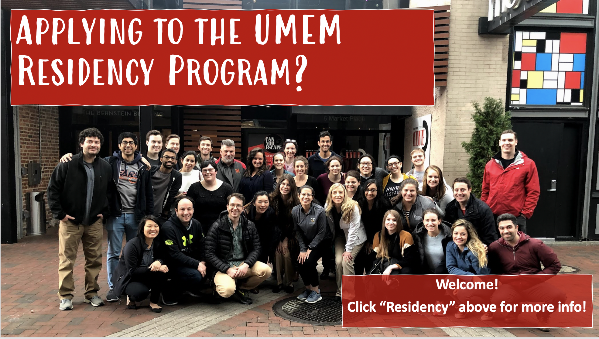
Welcome to the Department of Emergency Medicine at the University of Maryland School of Medicine.
We aim to be the preeminent academic department of emergency medicine, renowned globally for unparalleled education, unrivaled patient-centered care, pioneering research, and for training the next generation of innovative leaders in medicine.
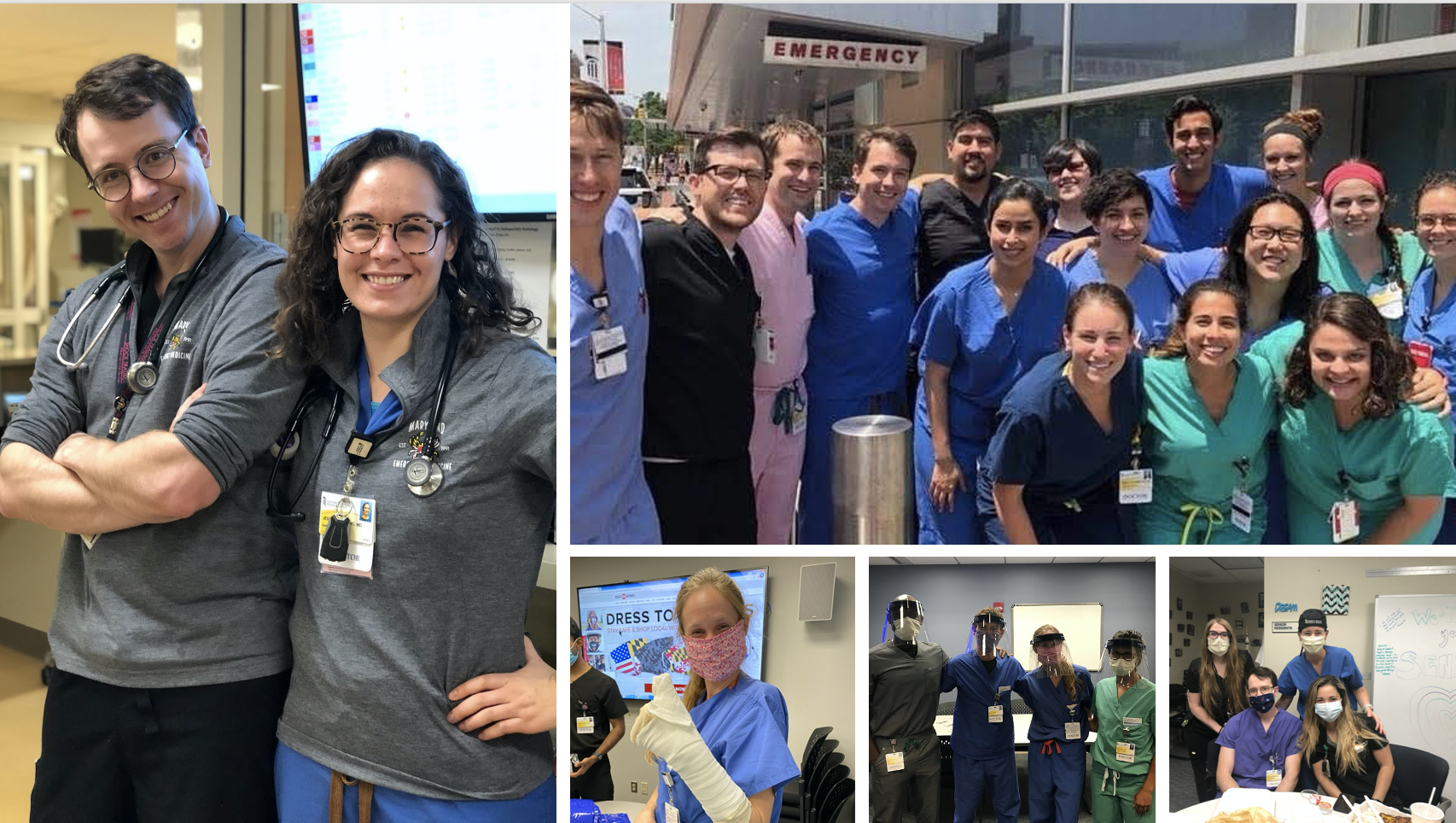
We are a multifaceted team of more than 80 faculty physicians shaping the future of emergency medicine. We teach at the bedside, in the classroom, and alongside colleagues at the local, national, and international levels. Over four decades, we have developed a well-earned reputation as top emergency medicine educators. Our faculty are thought leaders who publish textbooks and articles in leading emergency medicine journals, lecture nationally and internationally, and produce web-based materials that are trusted by countless physicians around the world.
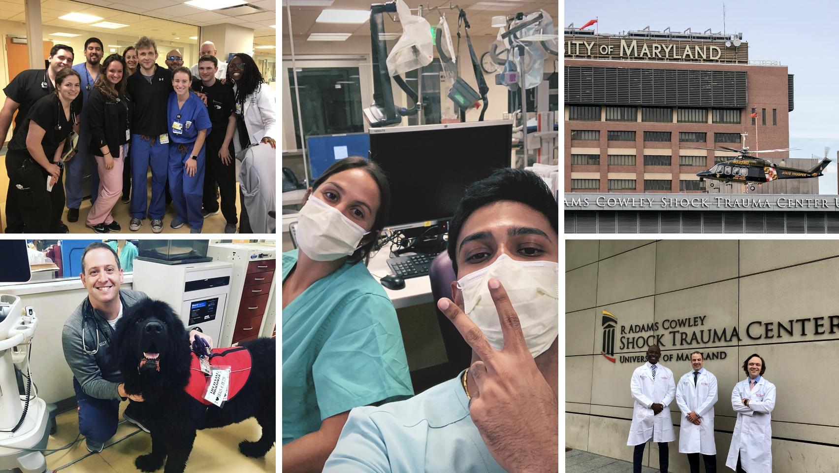
Every year, we treat more than 175,000 patients across the city of Baltimore—including at the University of Maryland Medical Center’s downtown and midtown campuses, Mercy Medical Center, and the Baltimore VA Medical Center. These facilities are enriched with state-of-the-art technology—from point-of-care ultrasound to hyperbaric chambers, advanced radiological and airway equipment, comprehensive stroke centers, and much more.
In addition to our highly regarded emergency medicine residency program, we host unique, combined multi-specialty training programs with internal medicine, internal medicine/critical care, and pediatrics. Our department also offers postgraduate fellowship training in Faculty Development, Simulation, Risk Management, Emergency Cardiology, Ultrasound, Administration, and Health Policy.
At the University of Maryland School of Medicine, our emergency medicine faculty are physicians—and so much more. We are educators, researchers, and healthcare administrators. We are entrepreneurs, policy experts, and medical informaticists. We are mentors, colleagues, leaders, and friends.
We are honored to serve our neighbors in Baltimore and the patients who trust us in their greatest times of need. Thank you for taking part in our dynamic mission to stabilize the injured, heal the sick, and save lives.
Mike Winters, MD, MBA
Chair, Department of Emergency Medicine
|
On Wednesday, June 17, faculty, residents, and staff from the University of Maryland School of Medicine’s Department of Emergency Medicine organized more than 250 hygiene kits for neighbors in need in the Baltimore community. (continued)
On June 10, 2026, the Department hosted its annual Research Day—an opportunity to highlight the breadth of Emergency Medicine research happening in the Department and elsewhere. (continued)
On June 12, 2026, the University of Maryland School of Medicine announced that Michael Winters, MD, MBA, will permanently serve as Chair of the Department of Emergency Medicine. (continued)
Should patients with mild cognitive impaired take glucosamine for their knee pain?... (continued)
This review article in the Journal of Trauma and Acute Care Surgery states: “In obtunded patients, an adequate and normal high-quality CT supports collar removal without the need for adjunctive imaging.”... (continued)
The first step toward developing a healthy ED is to develop a vision of the way emergency medicine should be practiced. The second is to analyze the way... (continued)
BLUF: Favor aggressive calcium supplementation following trauma, especially when patient requires transfusion. Recent evidence is pointing towards a signal... (continued)
Bottom Line: Among hospitalized patients boarding in the emergency department, care at an academic safety-net hospital, overnight admission, and elevated... (continued)