Vasopressor Tips in the Critically Ill
Head Impact Exposure and Concussion Incidence
There has been a major focus on head impact biomechanics as a cause of single-impact concussion in football.
The role of repeated subclinical (without diagnosed concussion) head impact exposure (HIE)
during the preseason and regular season may also be contributory.
There may exist individualized concussion tolerance levels. This threshold may be reduced by the burden of sustained subconcussive impacts
NCAA Division 1 football athletes sustain a median of 426 impacts over the course of a football season
652 impacts/season in high school football
Total head impact exposure during the preseason occurred at 2x the rate of the regular season
This association was investigated over 1120 athlete seasons from 6 NCAA D1 football programs across 5 years
Head Impact Telemetry was used to record head impact exposure
Elevated preseason HIE was strongly associated with preseason and in season concussion incidence
Total season HIE was strongly associated with total season concussion incidence.
Conclusion: There is a prolonged effect of HIE on concussion risk starting with preseason football.
Athletes with higher preseason HIE may have higher risk of concussion for the entire fall season.
In Practice:
In 2016, the Ivy League eliminated full contact practices from the regular season in addition to their existing limits on the amount of full contact in practice during the spring and preseason.
Currently, the NCAA has the following limitations: Teams won’t be allowed to hold full-contact practices on more than two days in a row. Each practice session is limited to only 75 minutes of full contact, in addition to a limit of two preseason scrimmages.
Enthusiasm for early transfusion of blood products in patients with traumatic shock has increased with increasing availability of pre-hospital blood and plasma and results of studies such as the PAMPer trial of pre-hospital plasma have shown potential mortality benefits. The deployment of prehospital blood for patients in hemorrhagic shock is promising but has significant cost and logistical considerations.
The RePHILL trial was a UK pre-hospital-based study of packed red blood cells and lyophilized plasma versus normal saline in trauma patients with presumed hemorrhagic shock. Patients older than age 16 with an SBP<90 or an absent radial pulse were eligible to get up to 1L of the study intervention. Multiple centers took part in the trial with 1:1 randomization stratified by study center. The primary outcome was a combination of mortality or lactate clearance less than 20% per hour or both.
A total of 432 patients were assigned a study fluid. The population was 82% male, median of 38 years old, with 78% of injuries classified as blunt, and 82% of the presumed hemorrhage classified at non-compressible. This was a very ill population with an average SBP of 73, an average GCS of 7 and an ISS of 36. The average from emergency call to EMS arrival was 30 minutes, average to study intervention was 26 minutes and time from EMS activation to ED arrival was 90 minutes.
The results showed no difference in the primary composite endpoint (64% vs 65%), with no difference in mortality (43% vs 45%) or lactate clearance (50% vs 55%). Interestingly, patients in the blood product arm had similar vital signs, lactate, and INR on ED arrival but received more blood products in the first 24 hours after ED arrival (pRBC 6.34 vs 4.41, p=0.004 and Plasma 5.04 vs 3.37, p=0.002). The was a trend toward improved early mortality at 3hr in the pre-hospital blood group (16% vs 22%, p=0.08).
Bottom Line(s):
Prehospital packed red blood cells and lyophilized plasma as compared to saline for traumatic shock did not improve mortality or lactate clearance in a well conducted multicenter RCT.
The use of prehospital blood products is promising but population which benefits, and the optimal type of product and delivery mechanism remain unclear.
Increased blood utilization and lower early mortality in the blood product group may represent alteration in the spectrum of disease that requires different early management.
The reasons for this counterintuitive result are unclear and further trials of whole blood as well as fibrinogen concentrates are ongoing.
Small Bowel Obstruction
Background:
Lung-protective ventilation with low-tidal volume improves outcome among patients with Acute Respiratory Distress Syndrome. The use of low tidal volume ventilation in the Emergency Departments has been shown to provide early benefits for critically ill patients.
Methodology:
A systemic review and meta-analysis of studies comparing outcomes of patients receiving low tidal volume ventilation vs. those who did not receive low tidal volume ventilation.
The authors identified 11 studies with approximately 11000 patients. The studies were mostly observational studies and there was no randomized trials.
The authors included 10 studies in the analysis, after excluding a single study that suggested Non-low tidal volume ventilation was associated with higher mortality than low tidal volume ventilation (1).
Results:
Comparing to those with NON-Low tidal volume ventilation in ED, patients with Low-Tidal volume ventilation in ED were associated with:
Discussion:
Conclusion:
Although there was low quality of evidence for low tidal volume ventilation in the ED, Emergency clinicians should continue to consider this strategy.
28-year-old male present with dorsal hand pain after “losing his temper”
On exam, you note dorsal swelling, tenderness, and deformity
AP, lateral and oblique views are obtained.
There is no rotational deformity but using the lateral view, you note that there is angulation
Measured as the shaft of the metacarpal as compared to the mid-point of the fracture fragment
Acceptable shaft angulation generally accepted to be less than 40°
Patient has greater that acceptable angulation so you have to perform closed reduction
After appropriate pain control consider the “90-90 method.”
Flex the MCP, DIP, and PIP joints to 90 degrees.
This positioning stretches the MCP collateral ligaments helping to optimize reduction
Next, apply volar pressure over the dorsal aspect of the fracture site while applying pressure axially to the flexed PIP joint.
Best demonstrated below
https://www.youtube.com/watch?v=40irKoUJqsM
-If the patient is able to maintain mentation/airway/SpO2/hemodynamics and cough up blood, intubation is not immediately necessary
-If you do intubate, intubate with the largest ETT possibly to faciliate bronchoscopic interventions and clearance of blood
-The CT scan that typically needs to be ordered is a CTA (not CTPA) with IV con
-See if you can find prior/recent imaging in the immediate setting (e.g. pre-existing mass/cavitation on R/L/upper/lower lobes)
-Get these meds ready before the bronchoscopist gets to the bedside to expedite care:
-If the pt's vent suddenly has new high peak pressures or decreased volumes after placement of endobronchial blocker, be concerned that the blocker has migrated
Encountered a situation in CCRU where we needed to prepare for a patient exsanguinating from gastric varices, and found a great summary of the different types of gastroesophageal balloons from EMRAP.
Summary: https://www.youtube.com/watch?v=Yv4muh0hX7Y
More in depth video on the Minnesota tube: https://www.youtube.com/watch?v=4FHIiA_doWU
Nice review article: https://www.sciencedirect.com/science/article/abs/pii/S0736467921009136
Aortic Dissection
Ultrasound has a great specificity for aortic dissection. Remember to take a look at your aorta on all cardiac views.
Let’s give a shout out to Nikki Cali for diagnosing aortic dissection in a patient with a recent PE. Can you find the dissection flap in this image?
Peritonsillar Abscess
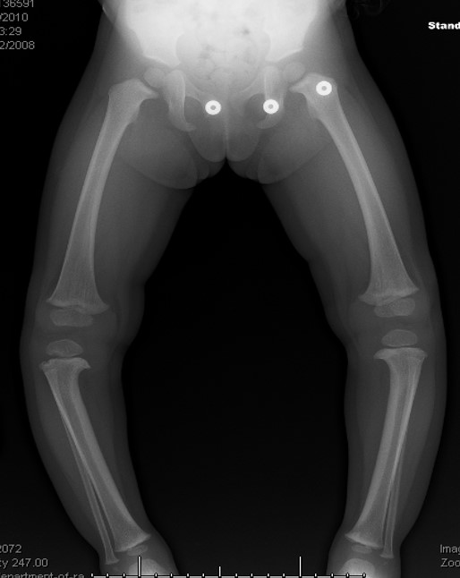
4-year-old patient comes to the ED for an unrelated complaint and you notice that his knees appear to be touching while his ankles remain apart.
Genu Varum or “knock knees” may be caused by Infantile Blount’s disease
-A progressive pathologic condition causing genu varum in children between ages 2 to 5
- Centered at the tibia
-Bilateral in up to 80%
-More common in boys
-Leg length discrepancy
- Articular incongruity
Risk factors: Early walkers (<1 year), overweight, large stature, Hispanic and African American
Results in disruption of normal cartilage growth at the MEDIAL aspect of the proximal tibia while LATERAL growth continues normally
May complain of knee soreness or subjective instability
On physical exam
Focal angulation of the proximal tibia
Lateral thrust during stance phase of walking (brief lateral shift of proximal fibula and tibia)
No tenderness or effusion
Imaging: Plain film shows varus deformity of the proximal tibia with medial beaking (beak like appears of bone) and downward slope of the proximal tibia metaphysis (increased metaphyseal-diaphyseal angle)
https://paleyinstitute.org/wp-content/uploads/blounts1.jpg
Treatment depends upon the age of the child and the severity
Successful in up to 80%
Note: In adolescent variant bracing is ineffective and surgery is only treatment
: Genu varum is normal in children <2 years old and becomes neutral at 14 months
DDX: Physiologic varus, Rickets


Appendicitis
Ultrasound has a reported high specificity (97.9) for acute appendicitis in moderate to high pre-test probability of patients.
Let’s give a shout out to Reed Macy, who diagnosed appendicitis in a male with vomiting and abdominal pain!
A recent prospective cohort study investigated the effect of low-dose droperidol on QTc in an emergency department:
Low-dose droperidol has a small effect on QTc and most patients remained below 500 ms.
Based on prior studies1 indicating possibly improved outcomes with vasopressin and steroids in IHCA (Vasopressin, Steroids, and Epi, Oh my! A new cocktail for cardiac arrest?), the VAM-IHCA trial2 compared the addition of both methylprednisolone and vasopressin to normal saline placebo, given with standard epinephrine resuscitation during in hospital cardiac arrest (IHCA).
The use of methylprednisolone plus vasopressin was associated with increased likelihood of ROSC: 42% intervention vs. 33% placebo, RR 1.3 (95% CI 1.03-1.63), risk difference 9.6% (95% CI 1.1-18.0%); p=0.03.
BUT there was no increased likelihood of favorable neurologic outcome (7.6% in both groups).
Recent publication on evaluation of long-term outcomes of the VAM-ICHA trial3 showed that, at 6-month and 1-year follow-up, there was no difference between groups in:
Bottom Line: Existing evidence does not currently support the use of methylprednisolone and vasopressin as routine code drugs for IHCA resuscitation.
9-year-old male left hand dominant, presents with left elbow pain.
He is a future “star pitcher,” says his coach dad. “Doc, I bet you didn’t know that although only 10% of people throw with their left hand almost a 1/3rd of MLB pitchers are lefties. He is 3x more likely than a righty to pitch in MLB.” “Maybe I’m asking him to throw too much.”
Hx: Lateral elbow pain and “stiffness” worse with activity that is better with rest
PE: Lateral elbow tenderness (capitellum) with slight (approx. 20 degrees) decreased loss of extension. Minimal swelling noted.
Dx: Panner's disease refers to osteochondrosis of the capitellum (similar to Legg Calve Perthes). Likely due to AVN from repetitive trauma. May also be due to endocrine disturbances.
Affects the dominant elbow of boys between the ages of 5 and 10
Associated with the repetitive trauma of throwing or gymnastics.
Must be differentiated from osteochondrosis dissecans which occurs in the older child >13yo when the ossification of the capitellum is complete
Radiology:
The articular surface of the capitellum may appear irregular or flattened with areas of radiolucency (43%). Loose bodies not seen with Panners, much more likely with OCD lesions.
Treatment: Ice and NSAIDs. Avoid pitching/gymnastics etc. until full radiographic and clinical healing. If significant pain and/or swelling place patient in long arm posterior splint for 7-10 days. Resolution may take several months and up to one year.
ED Low-Tidal Volume Ventilation
A recent study investigated the association between concussion and subsequent mental health conditions in a pediatric population.
Retrospective cohort study. Pediatric patients aged 5 to 18 years who presented to an ED, PCP or mental health practitioner from April 2010, to March 2020, in Ontario, Canada.
Primary outcome: Time to first diagnosis with a mental health condition during follow-up
Secondary outcomes: 1) self-harm 2) psychiatric hospitalization 3) death by suicide.
Mental health conditions: anxiety and neurotic disorders, adjustment reactions, behavioral disorders, mood and eating disorders, schizophrenia, substance use disorder, suicidal ideation, and disorders of psychological development.
Study group, almost 450,000 patients. Age and sex matching between those with concussion and those who experienced an orthopedic injury.
A significant association (P < .001) was found between concussion and mental health conditions
A significant association emerged between concussion and self-harm and psychiatric hospitalization
No association with suicide
Conclusion: Concussion was significantly associated with risk of mental illness, psychiatric hospitalization and self-harm but not death by suicide.
Concussed patients had an almost 40% higher rate of mental health conditions compared to controls (adjusted hazard ratio 1.39)
Take home: Screen patients who return to the ED with post concussive symptoms for mental health symptoms/concerns and provide appropriate awareness for parents


