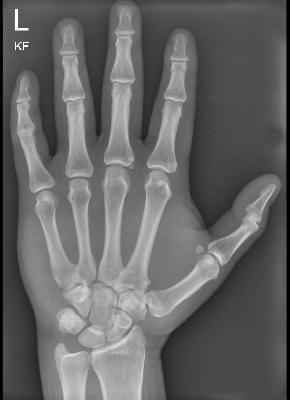
Pancreatic injury in pediatric blunt abdominal trauma is rare.
This study was a secondary analysis of the data collected by the Pediatric Emergency Care Applied Research Network (PECARN) in the Intra-abdominal Injury Study Group. Of the children who had blunt abdominal trauma, 6% had intraabdominal injuries and 1% had pancreatitis. A patient was considered as having traumatic pancreatitis if they had 2 of the following: 1) upper abdominal tenderness, 2) serum lipase of amylase > 3 x the upper limit of normal or 3) imaging study positive for pancreatitis.
Cervical spine injuries (CSI) are uncommon in children, but when present, they are often devastating. However, the application of a cervical collar in children is not benign and adverse effects include neck pain and discomfort and the downward tunnel vision it may create at the hospital leading to unnecessary testing. Recently, the PECARN group published a set of criteria to determine who requires imaging of the cervical spine in the emergency department. A planned subset of this initial study was to collect the impressions of EMS as they pertained to the 9 criteria to determine if these criteria could safely be used by EMS. The patients who were brought in by EMS with the potential for cervical spine injury, who were evaluated by the trauma team and/or had cervical imaging AND had electronic case reports filled out by EMS were included in the final analysis. Only 57% of the patients possible could be included based on the number of electronic case reports that were filled out.
Summary:
The recent ARISS (Albumin Resuscitation in Septic Shock) trial showed no difference in 90-day mortality or other secondary outcomes, similar to other trials comparing albumin and crystalloid. Notably however, the trial did not meet its predetermined enrollment requirement of patients (in the setting of the COVID-19 pandemic) and had a large portion of its intervention group failing to meet goal serum albumin level.
The Bottom Line:
There remains no evidence-based mortality benefit of albumin over crystalloid in patients with septic shock that do not have additional indications for albumin (such as hepatorenal syndrome). Crystalloid resuscitation remains a staple of appropriate and cost-effective care in septic shock. Albumin can be considered on a case-by-case basis after standard crystalloid resuscitation in this clinical setting.
This German retrospective study compared the prehospital use of ultrasound by trained paramedics and compared their findings to in-hospital diagnosis and image results. The authors found:
“Diagnostic accuracy, defined as the concordance between prehospital POCUS-based working diagnoses and final in-hospital diagnoses, was particularly strong for lung ultrasound (pneumothorax, pulmonary edema, pneumonia and pleural effusion; sensitivity 91.7%, specificity 100%) and eFAST (sensitivity 100%, specificity 96.5%), while for the abdominal ultrasound examinations, the specificity was 70% and sensitivity was 71.43%.”
This study sets the stage for future prospective work looking at prehospital US use by paramedics.
What elements of the history are most helpful for diagnosing a concussion?
An estimated 1.1 million to 1.9 million pediatric concussions occur annually in the US.
BOTTOM LINE:
A 2024 meta analysis from the European Society of Anaesthesiology and Intensive Care and British Journal of Anaesthesia worked to develop joint guidelines for best practices for intubation of neonates and infants.
While this guide is focused primarily on anesthesia and operative care, several of the recommendations have practical application to the EM and ICU as well.
They focused on general guidance including ensuring appropriate anesthesia and analgesia during intubation. But also discussed that videolaryngoscopy with standard blades is the most appropriate first line for all intubations in this age group. It allows for appropriate visualization either directly or by video and for learners allows instructors to observe as well.
When there are difficulties with intubation, hyperangulated blades have very high success rates, but LMA and video assisted intubation with a fiberoptic scope are also appropriate next steps for securing an airway.
When intubating, uncuffed endotracheal tubes are acceptable in all infants though cuffed are also safe in infants over 3kg in weight.
Finally, while apneic oxygenation is regularly used in adults, it is also recommended in the neonatal period to avoid hypoxia.
TenCRAOS was a phase 3 randomized, multi-center, double blind, double dummy, placebo-controlled trial in 78 patients that showed no significant difference in visual outcomes at 30 days for IV tenecteplase 0.25 mg/kg compared to aspirin 300 mg alone within 4.5 hours of central retinal artery occlusion (CRAO) symptom onset. Tenecteplase was associated with more serious adverse events, one of which was a fatal intracerebral hemorrhage.
Bottom line: Although tenecteplase has theoretical advantages in CRAO, the results of this trial do not support routine use.
This systematic literature review looking at gender differences in trauma care reveals:
In a large, randomized trial conducted in 42 ICUs in France, high-flow oxygen did not reduce 28-day all-cause mortality in adult patients with acute hypoxemic respiratory failure when compared to standard oxygen support.
A modified 2 round Delphi study was used to create 57 learning objectives in geriatric care for European prehospital providers. Based on in-hospital learning objectives and literature, these experts came up with what appears to be a very reasonable and helpful list of education objectives for pre-hospital providers that could easily apply to emergency medicine learners as a whole. Here is their table:
https://link.springer.com/article/10.1186/s13049-026-01550-3/tables/3
In this retrospective cohort study looking at splenic injury management and outcomes in the UK, patients over age 65 had much higher mortality and were more often managed conservatively (vs splenectomy or embolization) despite having a lower splenic injury grade and lower overall injury severity score compared to those under 65. Many factors are possible here including frailty, reluctance to intervene in older patients, and lower mechanism of injury bias away from evaluation and management.
Looking at trauma patients evaluated at a major trauma center before and after EMS switched from semi-rigid to soft cervical collars for immobilization found no difference in adverse outcomes. Add this to the mounting evidence that our current practice of spinal immobilization may not offer any benefit.

The 2026 Acute Pulmonary Embolism Guidelines recommend a new approach to risk stratification of patients with acute PE, including measurement of at least one cardiac biomarker and serum lactate, evaluation of RV size and function with CTA or echo (preferred when feasible), and multidisciplinary PERT assessment for all patients with acute PE and elevated clinical severity scores to assist with further risk stratification.

Bottom Line: Swimming-Induced Pulmonary Edema (SIPE) AKA Immersion Pulmonary Edema is a rare, though life-threatening pathology associated with water-based activities, especially among athletes or military personnel. Caused by physiologic effects of immersion, not from aspiration/ingestion. Consider in any patient with respiratory distress or chest discomfort onset during water activities such as swimming, diving, etc. Diagnose with physical exam and POCUS. Manage supportively, potentially including positive pressure ventilation. Screen for alternative diagnoses.
See the link for more thorough review of assessment diagnostics, pathophysiology, pharmacological options, risk factors, and long-term considerations.
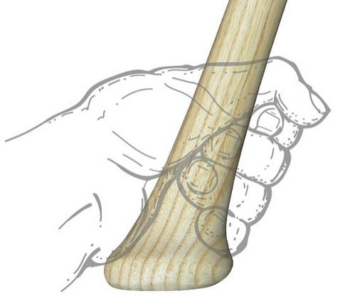
Bradycardia accompanying hypotension can be found in spinal cord injury (loss of autonomic reflex), beta blocker and calcium channel blocker overdose, intrinsic cardiac electrophysiologic derangement, and, often forgotten, intrabdominal hemorrhage. In the appropriate setting (blunt trauma, ruptured ectopic pregnancy), bradycardic hypotensive patients should be considered the same as tachycardic hypotensive patients and get a work up and treatment focused on Hemoperitoneum.
Using a database of 300,000 patients and applying a predictive measure for mortality, these authors found that patients over 66 with a high likelihood of 6 month mortality at the time of presentation were more likely to be admitted to an ICU when they presented to an ED. The authors conclude there is much work to be done regarding discussion of goals of care based on this information.
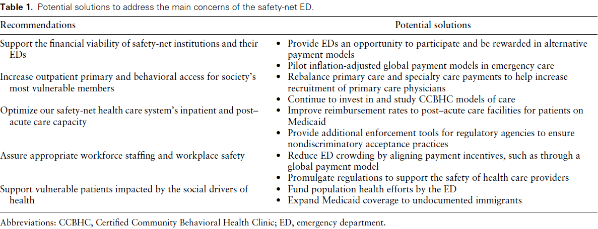
Bottom Line: Safety-net hospitals are those that see a substantial share of uninsured, Medicaid, or low-income Medicare patients. Their emergency departments (EDs) deliver disproportionally more undercompensated and uncompensated care, yet have similar operating costs as other EDs. Authors convened a group of 15 administrators of academic safety net EDs to identify and
develop a consensus understanding of barriers to delivering optimal care. See the link for details of their conclusions.
Click the link for below to read the bulleted, abridged version of the Executive Summary of the Updated SSC Guidelines for Adults with Sepsis and Septic Shock 2026…
