Wound Care:
Patients and many providers want to irrigate or wash a wound with an antiseptic solution in order to decrease the risk of infection. Most studies have shown that irrigation whether with tap water or sterile water is effective enough in reducing bacterial counts in a wound so does adding an antiseptic solution offer any additional benefit.
It turns out that hydrogen peroxide, and iodine based solutions can actually hinder wound healing as they causes delays in the migration and proliferation of fibroblasts at concentrations that are not even bactericidal. Chlorhexidine, and silver containing antiseptics [i.e.: silver sulfadiazine and silver nitrate] are bactericidal at concentrations that do not affect fibroblasts.
So in the end, if you feel the need to use an antiseptic, use chlorhexidine or a silver containing antiseptic. The use of hydrogen peroxide and iodine based solutions should be abandoned as they are not even bactericidal at concentrations that have profound affects on the fibroblasts.
There are a several classes of medications that can kill a toddler with a single dose. Toddlers are particularly susceptible due to their low weights and propensity to place everything in their mouths.
1. Calcium channel blockers
2. Camphor-containing rubs
3. Opioids/opiates
4. Oil of wintergreen/ aspirin
5. Cyclic antidepressants
6. Topical blood pressure patches (clonidine)
7. Eye drops and nasal sprays (oxymetazoline)
8. Sulfonylureas
9. Antimalarial drugs (cloroquine)
Fever is less common in infectious states in the elderly than in young patients. However, in contrast to younger patients, when an elderly patient does have a fever it is much more likely to be associated with a serious bacterial infection. It has been estimated that the source of fever in elderly ED patients is viral in only 5% of cases.
[from Hals G. Common diagnoses become difficult diagnoses when geriatric patients visit the emergency department, part I. Emergency Medicine Reports 2010;31(9):101-110.]
Septic Arthritis versus Arthritis:
Though CRP and ESR levels are significantly higher in patients that have septic arthritis, a 1998 study showed that there is extensive overlap between patients with septic arthritis crystal assoicated arthritis that both CRP and ESR have low sensitivity, specificity and predictive values. Peripherial WBC counts did not differ between the two disease processes..
The morale of the story: If you are suspecting septic arthritis you need to perform an arthorcentesis to analysis the synovial fluid. Systemic biomarkers can not support one diagnosis over the other.
some absolutes or almost always cases include the following:
Scombroid is caused by ingestion of preformed histamine on skin of fish.
Postcardiac Arrest Syndrome: Controlled Reoxygenation
Massive Pulmonary Embolism and Response to Fluids and Mechanical Ventilation
Massive pulmonary embolism leads to acute pulmonary hypertension and right ventricular overload. This leads to release of troponin and a "bowing" of the interventricular septum on echocardiography. Deviation of the septum then leads to a decrease in left-sided cardiac output.
A few interesting clinical pearls:
Elderly patients have slightly lower body temperatures than younger adults, and as a result it has been suggested that "fever" be defined as anything > 99 degrees F. One study found that by lowering the definition to this number improved the sensitivity and specificity to 83% and 89%, respectively.
from Hals G. Common diagnoses become difficult diagnoses when geriatric patients visit the emergency department, part I. Emergency Medicine Reports 2010;31(9):101-110.
study referred to: Castle SC, et al. Fever response in elderly nursing home residents: are the older truly colder? J Am Geriatric Soc 1991;39:853-857.
Osteomyelitis:
Hyperpronation: This reduction technique for a nursemaid's elbow (radial head subluxation) has been found to have better first attempt success than classic supination/flexion technique. (Pediatrics July '98). Support the elbow with a finger on the radial head, and forcefully hyperpronate.
Stroke strikes F.A.S.T. and must be recognized quickly for optimized management.
The following Face, Arms, Speech test, known as F.A.S.T., is an easy and quick bedside teaching tool that can be used to spread awareness about how to recognize and respond to stroke symptoms:
F = Ask person to smile. Does one side of face droop down?
A = Ask person to raise both arms. Does one arm drift downward?
S = Ask person to say a simple phrase. Does speech sound slurred or strange?
T = If any of the above findings are observed, it's time to call 911 immediately.
A single episode of hypotension portends a worse outcome for septic patients. The restrospective analysis by Marchick et al of 700 patients showed that mortality was 10% vs 3.6% for septic patients whose SBP dropped below 100 even once. It was also noted that the lower the SBP, the worse the in-hospital mortality.
So, not only do we need to remember to watch blood pressure closely for head-injured patients, but for septic patients as well!
We all know how difficult it can be to teach in the ED when it is busy. So how do the experts do it when there is so little time?
Just a few considerations that might make your teaching more effective and easier to do when it is busy:
Elderly patients should be considered immunocompromised for several reasons:
1. T cell function and reduced cellular immunity occur as we get older.
2. B cell antibody production decreases.
3. Host defenses against infection are reduced with aging, such as reduced circulation and thinning skin.
4. Miscellaneous factors, such as malnutrition and co-existing illnesses contribute to increased risk of infection as well.
[Good reference and suggested reading: Hals G. Common diagnoses become difficult diagnoses when geriatric patients visit the emergency department: Part I. Emergency Medicine Reports 2010;31(9):103-111.]
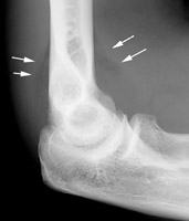
Radial Head Fractures:
Radial head fractures can often be difficult to visualize on plain films especialing Mason Type 1 fractures (see prior pearl on classification system) which are nondisplaced. Often the only sign of a fracture will be a posterior fat pad sign which is always considered to be pathologic. The posterior fat pad lies outside the synovium of the elbow joint and is normally hidden in the fossa of the distal humerus preventing it from being seen on lateral films of a normal elbow. Trauma to the elbow that results in a intraarticular fracture (typically a radial head fracture) produces an intra-articular hemorrhage that distends the synovium and displaces the fat out of the fossa, producing the typical triangular radiolucent shadow posterior to the distal end of the humerus.
Once you've made the presumptive diagnosis of cerebral edema in Pediatric DKA (refer to part 1), here's what's next:
Mortality from cerebral edema in DKA is 20-25%, and 15-35% of survivors have permanent disability.
The best strategy is to do your best to avoid cerebral edema in the first place, but if you do recognize it, this is a clinical diagnosis, and you should not delay treatment for radiographic studies.