UMEM Educational Pearls - Vascular

Category: Vascular

Title: Aortic Root Measurement

Keywords: aortic aneurysm; point-of-care ultrasound; pocus; aortic dissection (PubMed Search)

Posted: 12/4/2023 by Alexis Salerno, MD (Updated: 4/20/2024)
Click here to contact Alexis Salerno, MD

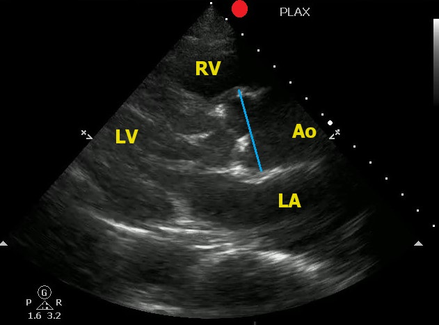
Point-of-Care Ultrasound can help to identify signs of thoracic aortic dissection.

One view to help in your assessment is the Parasternal Long Axis View.

  • The aortic root should be in a 1:1:1 ratio with the left atrium and the right ventricle.
  • The aortic root should be less than 4 cm (4.5 cm considered aneurysmal)

To correctly measure the aortic root:

  • Measure at the Sinus of Valsalva
  • Measure during diastole (when the aortic valve is closed)
  • Measure leading edge to leading edge

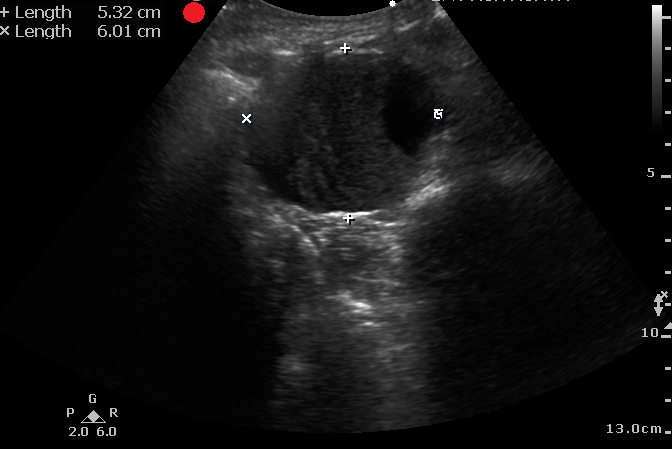
Here is an example of an aortic root aneurysm: 

Show References


Category: Vascular

Title: POCUS for Abdominal Aortic Aneurysm

Keywords: POCUS, AAA, (PubMed Search)

Posted: 6/19/2023 by Alexis Salerno, MD (Updated: 4/20/2024)
Click here to contact Alexis Salerno, MD

It is difficult to diagnosis a ruptured AAA with POCUS. However, based on one systematic review and meta-analysis, POCUS has a sensitivity of 97.8% and a specificity of 97% for diagnosing AAA in patients supsected of having a ruptured AAA. 

Remeber:

  • The normal abdominal aorta should measure under 3 cm.
  • The proximal iliacs should measure under 1.5 cm. 
  • Measure the aortic diameter from leading edge to leading edge. 

Laslty, make sure you are measuring the aortic wall and not a mural thrombus. 


Show References


Category: Vascular

Title: Complications of Acute Aortic Dissection

Keywords: Aortic Dissection (PubMed Search)

Posted: 6/20/2011 by Rob Rogers, MD (Updated: 4/20/2024)
Click here to contact Rob Rogers, MD

There are several complications of acute aortic dissection that emergency physicians must be familiar with.

These include:

  • Cardiac tamponade (most common cause of death)
  • Acute aortic regurgitation
  • Stroke
  • Free intrathoracic rupture
  • Malperfusion syndrome (kidney, spinal cord, bowel, extremity, etc.)

*Key Pearl: If a patient with suspected or confirmed acute aortic dissection suddenly arrests consider cardiac tamponade.

Show References


Category: Vascular

Title: Image of the Week

Posted: 1/3/2011 by Rob Rogers, MD (Updated: 4/20/2024)
Click here to contact Rob Rogers, MD


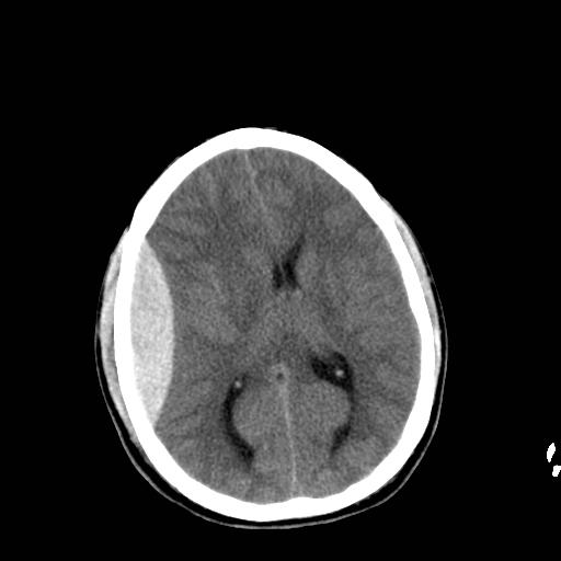
60 year-old male s/p assault. + LOC. Awake and normal neuro examination on arrival. Deteriorates in the ED after about an hour....

Diagnosis: Epidural Hematoma


Show References


Category: Vascular

Title: Diagnosing Subarachnoid Hemorrhage-6 Pitfalls

Keywords: subarachnoid hemorrhage (PubMed Search)

Posted: 12/13/2010 by Rob Rogers, MD (Updated: 4/20/2024)
Click here to contact Rob Rogers, MD

Diagnosing Subarachnoid Hemorrhage-6 Pitfalls

1. Subarachnoid hemorrhage (SAH) doesn't always present as the "worst ever" headache. Don't most of our patients say their headache is the worst headache anyway? Be suspicious of the diagnosis if your patient has acute onset of an unusual or atypical headache. Diagnoses starts with the history.

2. The neuro exam may be completely normal in some cases, especially early on.

3. The headache due to SAH may get better with analgesics. This is a huge pitfall. Don't rule this diagnosis out if analgesics help.

4. The CT scan may be negative. Enough said.

5. Be careful with interpretation of the CSF. We all want the number of red cells in tube 4 to be zero. Be careful with this. Although the rbcs may have dropped by 50% from tubes 1 to 4, the diagnosis hasn't been excluded unless the cells clear completely. Although there have been some case reports of SAH with rbcs < 100, this is pretty uncommon.

6. CT Angiography and/or MRI with FLAIR is not a substitute for the lumbar puncture.

Show References


Category: Vascular

Title: Fenoldopam (Corlopam) dosing

Keywords: hypertension (PubMed Search)

Posted: 12/6/2010 by Rob Rogers, MD (Updated: 4/20/2024)
Click here to contact Rob Rogers, MD

Got Symptomatic Hypertension? Why not try Fenoldopam?

Fenoldopam is a rapid-acting vasodilator.

  • It is an agonist for D1-like dopamine receptors and binds with moderate affinity to α2-adrenoceptors.
  • Effective as nitroprusside, however, it has the advantages of increasing renal blood flow (6 times as potent as dopamine in producing renal vasodilitation) and sodium excretion
  • Not associated with the accumulation of toxic metabolites, and not requiring shielding from light.
  • Fenoldopam can be safely used in all hypertensive emergencies, and may be particularly beneficial in patients with renal insufficiency.

Dosing (Adult): After a starting dose of 0.1 to 0.3 mcg/kg/minute, the dose is titrated at 15 minute intervals, depending on the BP response. May be increased in increments of 0.05 to 0.1 mcg/kg/minute every 15 minutes until target blood pressure is reached. Maximal infusion rate reported in clinical studies: 1.6 mcg/kg/minute.

Onset/duration: 5-10 minutes/~ 1 hour.

Show References


Category: Vascular

Title: Pulmonary Embolism and IVC Filters

Keywords: Pulmonary Embolism, IVC Filter (PubMed Search)

Posted: 9/20/2010 by Rob Rogers, MD (Updated: 4/20/2024)
Click here to contact Rob Rogers, MD

Pulmonary Embolism and IVC Filters

Inferior vena cava filters are placed in patients with massive DVT and /or in patients who cannot receive systemic anticoagulation.

The question is, can patients develop pulmonary embolism if a filter is already in place? The answer: yes

How does this happen?:

  • Clot burden at the site of cava-filter insertion (below the filter). Clots can dislodge at this site and slip through the filter.
  • Embolization around the IVC filter via retroperitoneal collaterals.


Category: Vascular

Title: Hypertensive Encephalopathy-Difficulty with Diagnosis

Keywords: Hypertension (PubMed Search)

Posted: 8/30/2010 by Rob Rogers, MD (Updated: 4/20/2024)
Click here to contact Rob Rogers, MD

Hypertensive Encephalopathy (HE) is a clinical diagnosis and can look like many other disease entities.

HE refers to a relatively rapidly evolving syndrome of severe hypertension in association with severe headache, nausea, and vomiting, visual disturbances, convulsions, altered mental status and, in advanced cases, stupor and coma.

The key is the presence of severe hypertension. Remember, though, that 160/105 mm Hg may be high for an individual patient. Most patients with the syndrome will have diastolic pressures well in excess of 120-130 mm Hg. The only way you will know if the diagnosis is correct is to treat the BP (carefully control), work up other etiologies, and see of symptoms improve with BP control.

Beware the patient with severe HTN and seizure. Seizure may be the first, and only, symptom of hypertensive encephalopathy. 


Category: Vascular

Title: Beta Blockade in Treating Acute Aortic Dissection

Keywords: Aortic Dissection (PubMed Search)

Posted: 8/23/2010 by Rob Rogers, MD (Updated: 4/20/2024)
Click here to contact Rob Rogers, MD

Beta Blockade in Treating Acute Aortic Dissection

Medical therapy for acute aortic dissection is aimed at decreasing shear stress within the aorta. Although there are many agents to choose from when treating hypertension in patients with acute aortic disease, all regimens should include a beta blocker (like esmolol) unless contraindicated. Initiation of a beta blocker before another antihypertensive agent is added is crucial as this will prevent reflex tachycardia associated with vasodilators and other afterload reducers. Reflex tachycardia may worsen the dissection. 


Treatment of Cerebral Venous and Sinus Thrombosis

Thrombosis of the cerebral venous system, also known as cerebral venous and sinus thrombosis and dural sinus thrombosis, is an uncommon condition encountered in the emergency department. The diagnosis may be stumbled upon by various CT findings or by MRI and/or a high opening pressure on lumbar puncture.

The treatment of choice is full dose anticoagulation with heparin. Available studies looked at unfractionated heparin, but many experts now consider LMWH (like Lovenox) an acceptable alternative. Despite the risk of hemorrhagic transformation of a venous infarct, heparin therapy is considered the standard treatment for this condition. 

Show References


Category: Vascular

Title: Pulmonary Embolism and Blood Pressure

Keywords: Pulmonary Embolism (PubMed Search)

Posted: 8/2/2010 by Rob Rogers, MD (Updated: 4/20/2024)
Click here to contact Rob Rogers, MD

Pulmonary Embolism and Blood Pressure

Patients with massive PE will often develop worsening hypotension after a fluid bolus due to increased right ventricular distension and deviation of the interventricular septum towards the left side of the heart. This septal deviation decreases left heart cardiac output.

In addition, patients with massive PE will sometimes develop higher blood pressures after intubation as positive pressure ventilation reduces preload, decreases deviation of the septum, and increases left sided cardiac output.


Category: Vascular

Title: Silent Pulmonary Embolism

Keywords: Pulmonary Embolism (PubMed Search)

Posted: 6/28/2010 by Rob Rogers, MD (Updated: 4/20/2024)
Click here to contact Rob Rogers, MD

Silent Pulmonary Embolism?

As many as 50% of patients with isolated DVT will be found to have silent pulmonary embolism (i.e. no chest pain or shortness of breath) on VQ scanning. Studies performed in the last year or so with CT scanning show that this percentage is much higher.

The clinical take-home point is NOT to get a pulmonary CTA on suspected DVT patients but to remember that many patients can and do have PE in the absence of cardiopulmonary symptoms. Pretty frightening....

Show References


Category: Vascular

Title: Got Lytics?

Keywords: Pulmonary Embolism (PubMed Search)

Posted: 6/7/2010 by Rob Rogers, MD (Updated: 4/20/2024)
Click here to contact Rob Rogers, MD

Thrombolytic Therapy for Pulmonary Embolism

Current, FDA-approved thrombolytic therapy for PE:

  • tPA 100 mg over two hours-infusion
  • Heparin drip should be turned off during tPA infusion and turned back on ONLY after PTT has fallen to 2 X normal
  • Other drugs are being used-like Tenecteplase (TNKase), but strictly speaking, not FDA approved for thrombolysis of PE
  • Most studies to date do not show that catheter-based delivery of lytics is safer than systemically administered lytics


Category: Vascular

Title: Massive Pulmonary Embolism and Response to Fluids

Keywords: Pulmonary Embolism (PubMed Search)

Posted: 5/24/2010 by Rob Rogers, MD (Updated: 4/20/2024)
Click here to contact Rob Rogers, MD

Massive Pulmonary Embolism and Response to Fluids and Mechanical Ventilation

Massive pulmonary embolism leads to acute pulmonary hypertension and right ventricular overload. This leads to release of troponin and a "bowing" of the interventricular septum on echocardiography. Deviation of the septum then leads to a decrease in left-sided cardiac output. 

A few interesting clinical pearls:

  • Administration of IV fluids to patients with massive PE often leads to a decrease in BP. This happens as a result of increasing preload causing further bowing of the septum and a subsequent further drop in left ventricular cardiac output, leading to hypotension. 
  • Patients with massive PE who require intubation often demonstrate an increase in BP due to positive pressure ventilation causing a drop in preload and a reduction of septal bowing into the left ventricle.


Category: Vascular

Title: More on the PERC rule

Keywords: PERC, pulmonary embolism (PubMed Search)

Posted: 4/26/2010 by Rob Rogers, MD (Updated: 4/20/2024)
Click here to contact Rob Rogers, MD

A review of the PERC rule...

The "PERC Rule"  is used to assess a patient's risk for probability of PE in the emergency department. It involves evaluating the presence or absence of 8 clinical criteria to arrive at a pretest probability.  And remember, this rule is supposed to be used for patients with really low pretest probability where you weren't concerned about PE to begin with. Some experts claim that "PERC negative" on the chart proves you considered PE in the differential diagnosis. But the test isn't designed to be used on EVERY patient as a means to rule out PE. Only use if you thought about the disease in a low risk patient and didn't plan on getting a d-dimer or further testing. 

The criteria are (all must be YES):


age < 50 years

heart rate less than 100 beats per minute

room air oxygen saturations 95% or greater

no prior deep venous thrombosis [DVT] or PE

no recent trauma or surgery (4 weeks)

no hemoptysis

no exogenous estrogen

no clinical signs suggestive of DVT (Unilateral leg swelling on visual inspection

Show References


Category: Vascular

Title: Secondary Hypertension...Say What?

Keywords: Hypertension (PubMed Search)

Posted: 4/19/2010 by Rob Rogers, MD (Updated: 4/20/2024)
Click here to contact Rob Rogers, MD

Secondary Hypertension...say what?

We obviously see tons of patients in the ED with hypertension, and we are very comfortable with both symptomatic and asymptomatic presentations. Most of these patients have essential or primary hypertension. Some patients, however, may have secondary hypertension (i.e. something is causing it). Although we will refer patients to a primary care physician for further management and workup it is worth discussing when to suspect other diagnoses as the cause of the hypertension. Is it out job necessarily to diagnose these conditions in the ED? No. 

Causes of secondary hypertension to consider:

  • Obstructive sleep apnea
  • Renal disease
  • Renal artery stenosis (think older person with HTN and abdominal bruit)
  • Coarctation (young person with HTN-ever wonder why pediatricians palpate upper and lower extremity pulses in the office?)
  • Cushing's disease (excess cortisol-patient may have new diabetes, have abdominal striae, and easy bruising)
  • Hyperaldosteronism (due to an adrenal tumor)...think about if a patient comes to the ED and is repeatedly hypokalemic and hypertensive
  • Pheochromocytoma (episodes of flushing, hypertension, palpitations, etc.)
  • Hypothyroidism (not myxedema coma or storm)...commonly causes elevated diastolic BP. 
  • Hyperthyroidism 

Consider the ABCDE mnemonic:

A-Accuracy (is it really htn?), Apnea, Aldosteronism

B-Bruits, Bad Kidneys

C-Catecholamines, Coarctation, Cushing's 

D-Drugs, Diet

E-Endocrine


Aren't you glad you didn't do a Medicine residency???

Show References


Category: Vascular

Title: Type B (distal) Aortic Dissection-Beware of Complications!!

Keywords: aortic dissection (PubMed Search)

Posted: 4/5/2010 by Rob Rogers, MD (Updated: 4/20/2024)
Click here to contact Rob Rogers, MD

Some not too uncommon complications of Type B (distal) aortic dissection:

  • Malperfusion syndrome-occurs when the dissection flap occludes a major vessel (e.g. SMA occlusion leading to bowel infarction)
  • Occlusion of the spinal arteries and lower extremity arteries can lead to fleeting signs and symptoms-one minute they have left leg pain and ischemia, the next minute they don't. This is pretty classic for acute, distal aortic dissection. 
  • Frank rupture (dissected aortic wall is weak and prone to aneurysm formation and subsequent rupture)
  • Assume that rupture has occurred (may be intrathoracic or intrabdominal) in a Type B patient who crashes unexpectedly
  • Retrograde extension into the proximal aorta is not common but does occur. Have a low threshold to whip out the sono if the patient deteriorates. 


Category: Vascular

Title: Acute Mesenteric Ischemia

Posted: 3/29/2010 by Rob Rogers, MD (Updated: 4/20/2024)
Click here to contact Rob Rogers, MD

Acute Mesenteric Ischemia

Although we all know the classic presentation of acute mesenteric ischemia (AMI), it can be tough to diagnose.

Some pearls about AMI:

  • Embolization to the superior mesenteric artery (SMA) is the most frequent cause of AMI.
  • Most patients present with acute, severe abdominal pain. 
  • Classic presentation: acute severe abdominal pain with a paucity of physical examination findings
  • Presence of tenderness in most cases indicates bowel infarction has already occurred
  • The disease may be more insidious in patients with diseased mesenteric vessels (presence of collaterals). These patients may very well NOT present with acute, severe pain.
  • Must have a high index of suspicion (i.e.-suspect this disease in patients at risk who present with abdominal pain)
  • If you are standing at the bedside and you say, "Self, this looks like AMI," then rally the troops BEFORE labs and before CT. Get a surgeon to see the patient as soon as possible. Tell them you think the patient has AMI. Get them to move. "TIme is bowel."


Category: Vascular

Title: Splenic Artery Aneurysm

Keywords: Aneurysm (PubMed Search)

Posted: 3/8/2010 by Rob Rogers, MD (Updated: 4/20/2024)
Click here to contact Rob Rogers, MD

Splenic Artery Aneurysm (SAA)

Ever scanned someone and the report says "incidental note of a splenic artery aneurysm"? Well, if it hasn't happened yet, it will sooner or later. This type of aneurysm isn't that rare and with the number of abdominal CTs we order we are bound to see this in clinical practice.

Some important points to remember about SAA:

  • 3rd most common location of intra-abdominal aneurysm, 1st-aortic aneurysm, 2nd-iliac artery aneurysm
  • Most common complication is sudden rupture and occurs in as many as 3-10% of cases
  • 80% pf patients with SAA are asymptomatic
  • Symptomatic aneurysms may present with left upper quadrant pain, nausea, and vomiting
  • Splenic infarct is a rare complication
  • Most important is followup: patients will need close followup for asymptomatic splenic artery aneurysms. Consultation with a surgeon will need to be arranged if it is thought that the patient has symptoms due to the aneurysm


Category: Vascular

Title: Suspect Aortic Dissection-Don't Wait to Start the Drip!

Keywords: Aortic Dissection (PubMed Search)

Posted: 2/22/2010 by Rob Rogers, MD (Updated: 4/20/2024)
Click here to contact Rob Rogers, MD

Suspect your patient has an aortic dissection? Don't wait to lower the blood pressure.

A few considerations for the patient with suspected aortic dissection:

  • If the patient is hypertensive AND you really think they could have the disease, start the drip then. Don't wait until the CT is done. Every second that goes by with extremely elevated BP may increase the dissection length.
  • If you are really worried about a patient having a proximal aortic dissection, don't wait for the creatinine to come back...scan them without it. If you are really suspicious this is justified in many cases.
  • There is very little (to no) role in performing a dry CT (because the patient's creatinine comes back elevated). Dry CT is very insensitive. Instead get a transesophageal echo or an MRI.