Many patients present to the emergency department for ankle swelling. On way to identify signs of intra-articular swelling is to use POCUS. To perform this, place the linear probe at the tibio-talar junction with the probe marker placed towards the patient’s head. An effusion is identified as anechoic fluid in-between the tibia and talus bone.

POCUS has been shown to improve first-pass success and overall success as compared to a landmark based approach for medium-sized joints. When performing an ankle arthrocentesis with POCUS, care should be taken to avoid blood vessels and tendons.
What are the signs of Cardiac Tamponade on ultrasound?
Think of them as a pyramid with clinical importance decreasing as you rise to the top of the pyramid.
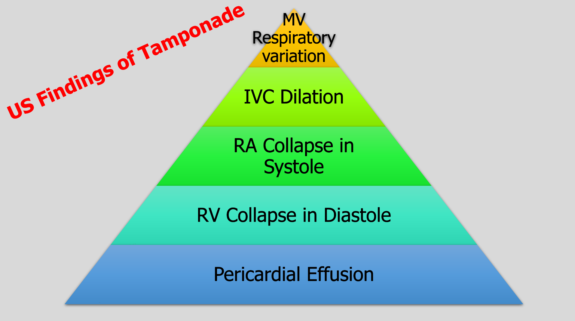
To have tamponade you need a pericardial effusion.
The most specific sign of tamponade is RV collapse in diastole.
The earliest and most sensitive sign is RA collapse over 1/3 of the cardiac cycle from late diastole into systole, which is why we say RA collapse during systole.
IVC dilation also occurs but is not sensitive.
Placing the pulse wave Doppler over the mitral valve and evaluating the change with respirations is an advanced technique. It’s positive if you have 25% change.
Don’t know if you are in systole or diastole? Connect your telemetry leads to the ultrasound machine. Don't have leads? Then you can also cine scroll on a subxiphoid view or parasternal view to look at when the valves are open and closed, then compare to the cardiac wall positioning.
By performing a Point-of-Care Transvaginal Ultrasound (TVUS), we can decrease length of stay for patients with early pregnancy. Moreover, if an ectopic pregnancy is identified, we can decrease time to the OR for these patients.
Begin by discussing the exam with the patient and ensuring they have emptied their bladder. Apply a probe cover and add sterile lubricant to the outside of the probe tip. You can save time by performing a TVUS immediately after the pelvic speculum exam for swab collection.
Gently introduce the transducer with the marker upward, directed towards the ceiling. As you slowly advance, the uterus will be visualized in a sagittal orientation. Fan through the uterus by moving the probe handle left and right.
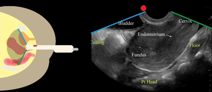
Image From: doi: 10.1016/j.emc.2022.12.006.
Rotate the transducer so that the marker is directed towards the patient's right side. Fan through the uterus by lifting the probe handle up and down.
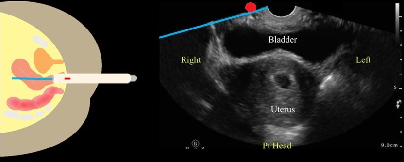
Image From: doi: 10.1016/j.emc.2022.12.006.
If a gestational sac is found, you should measure the gestational age and if present, fetal heart rate.
Tilt the transducer towards the patient's left or right side to visualize the adnexa. The adnexa will be located medially to the iliac vessels.
Remove the transducer and follow your department protocol for high level disinfection.
Point-of-Care Ultrasound can help to identify signs of thoracic aortic dissection.
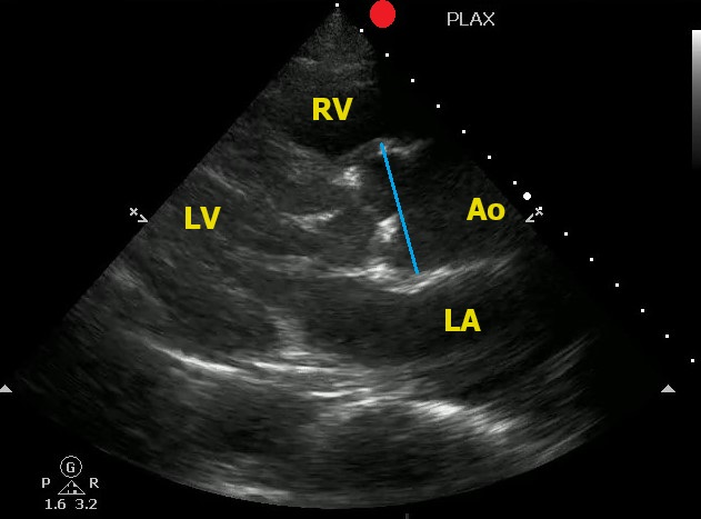
One view to help in your assessment is the Parasternal Long Axis View.
To correctly measure the aortic root:
Here is an example of an aortic root aneurysm:
Acute bronchiolitis (AB) is a common cause of respiratory tract infections in infants. A recent study looked at the application of Point-of-Care Lung Ultrasound (LUS) in infants <12 months who presented with symptoms of AB.
They scored infant lungs using a cumulative 12-zone system. With the below scale:
0 - A lines with <3 B lines per lung segment.
1 - ≥3 B lines per lung segment, but not consolidated.
2 - consolidated B lines, but no subpleural consolidation.
3 - subpleural consolidation with any findings scoring 1 or 2.
They found that infants with higher LUS scores had increased rates of hospitalization and length of stay.
Here are some tips for ultrasounding a pediatric patient:
Point of Care Ultrasound has been shown to change medical management and decrease time to diagnosis.
However, sometimes on a busy shift we may get an xray or radiological study prior to performing a POCUS exam due to time constraints.
A recent study looked at the time it takes to perform a bedside ultrasound.
The authors measured the duration of time from starting the exam through the ultrasound worklist to the timestamp on the last recorded image.
They reviewed 2144 studies and found a median time of 6 minutes to perform a study.
Of course the study is limited by the time it takes to find a machine, make sure it is functioning and other supplies such as gel.
Conclusion: You can take 6 minutes to assist in your patient's clinical care.
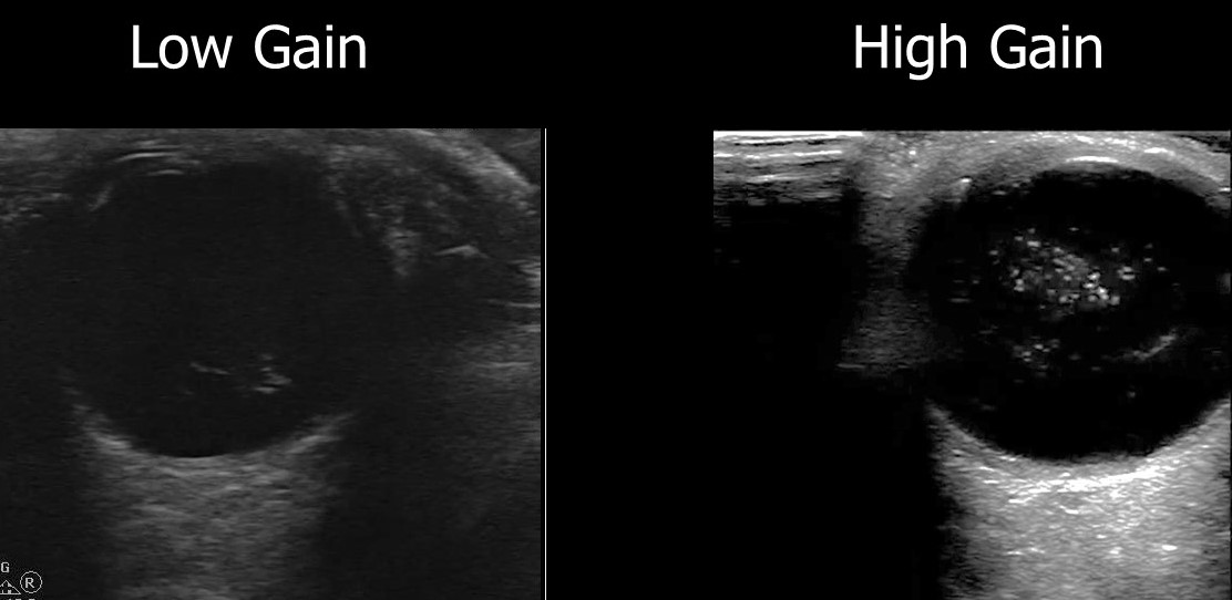
Prior research has shown that EPs can accurately detect ocular pathology using POCUS.
A recent retrospective review looked at how ultrasound image gain levels impacted the accuracy of POCUS for detection of retinal detachment, retinal hemorrhage and posterior vitreous detachment.
They included 383 ED patients who received ocular POCUS and ophthalmology consultations.
Conclusions: The authors found that increasing the gain for ocular POCUS images allowed for increased sensitivity.
Here is an example of a vitreous hemorrhage on ocular POCUS using low gain and high gain.
Ectopic pregnancy ranges from 3 to 13% in symptomatic first-trimester ED patients.
The discriminatory zone is defined as the level of Bhcg above which an intrauterine pregnancy can be reliably detected using ultrasound. (1,500 mIU/mL for transvaginal ultrasound and 3,000 mIU/mL for transabdominal ultrasound)
One study found that an intrauterine pregnancy was visualized with as low as 1,440 mIU/mL and patients with an interdeterminate pelvic ultrasound who were found to have an ectopic pregnancy had a Bhcg greater than 3,000 mIU/mL only 35% of the time.
Bottom Line: If you have a symptomatic patient with an empty uterus and a bhcg above the discriminatory zone, they have a higher risk for ectopic pregnancy. However, if your patient is symptomatic, they should still have further evaluation for ectopic pregnancy even if they have a bhcg lower than the discriminatory zone.
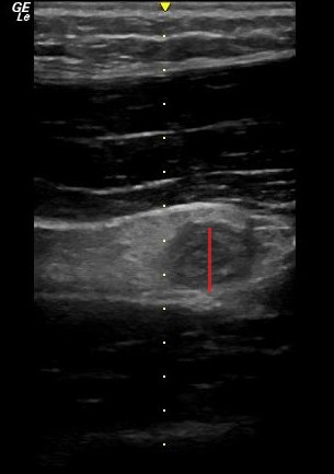
POCUS can be used to screen for appendicitis.
A recent study showed a sensitivity of 66.7% (CI 95% 47.1–82.7), and a specificity of 96.8% (CI 95% 83.3–99.9) during pregnancy, with the highest sensitivity in the first trimester.
2 methods to locate the appendix are:
1) have your patient point to the area where it hurts the most
2) perform a lawnmower technique over the right lower quadrant looking for the right psoas mucle and the iliac vessels. The appendix will usually be near these structures.
Sometimes it is easiest to use your curvilinear probe to identify an area of inflammation and then change to the linear probe for better visualization.
On ultrasound, appendicitis is defined as a non-compressible blind pouch with an outer diameter greater than 6 mm. On short axis the inflammed appendix will look like a target sign:
The use of a fascia iliaca compartment block has been shown to reduce pain, decrease length of stay and decrease the opiate requirements for patients with hip fractures.
Check out this page on how to perform this procedure.
Fascia iliac blocks can be challenging to implement routinely in the emergency department. Studies show that 2.5% of eligible patients, despite departmental implementation, receive a block.
One recently published article showed that large scale multi-disciplinary implementation can increase the use of fascia iliac blocks. After implementation, the study team found that 54% of eligible patients received a fascia iliac block.
This article is interesting as it provides helpful resources including physician and nursing protocols for performing this block.
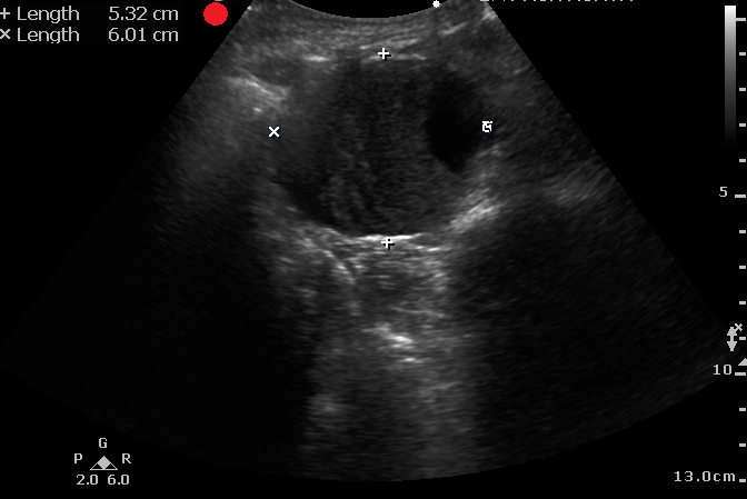
It is difficult to diagnosis a ruptured AAA with POCUS. However, based on one systematic review and meta-analysis, POCUS has a sensitivity of 97.8% and a specificity of 97% for diagnosing AAA in patients supsected of having a ruptured AAA.
Remeber:
Laslty, make sure you are measuring the aortic wall and not a mural thrombus.
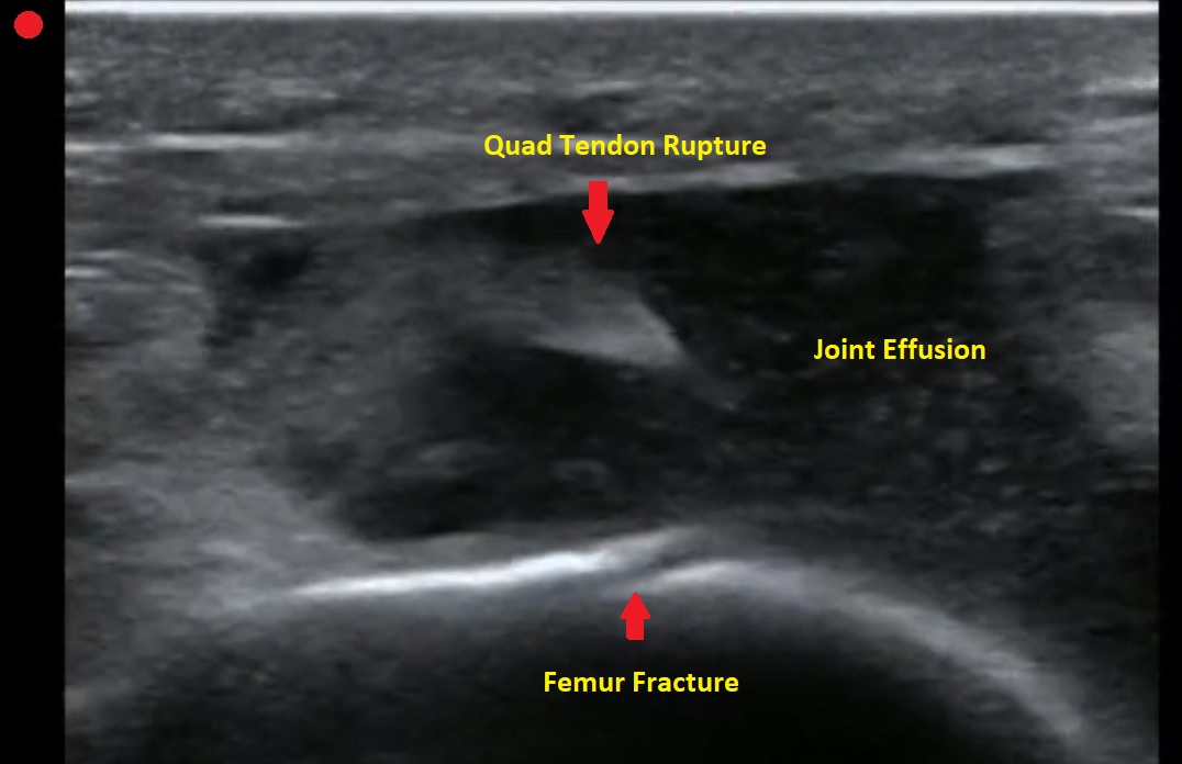
Pt presents to the emergency department with knee pain.
You decide to ultrasound the proximal knee. You place your ultrasound probe in the midline of the knee with your probe marker towards the patient's head.
What is the diagnosis?
-
--
---
--
-
The answer is a quadriceps tendon rupture with femur fracture.
We hope that you enjoy your Memorial Day!
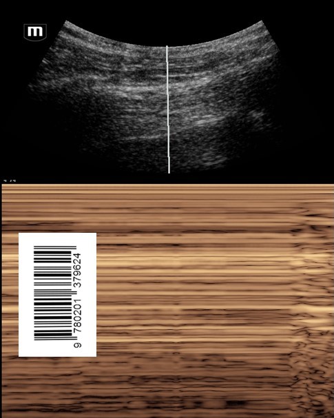
Don't forget your Sandy Beach Sign vs Barcode Sign of Lung Ultrasound:
Normal lung will have good pleural sliding. When you image the lung with M-Mode it looks like a Sandy Beach.
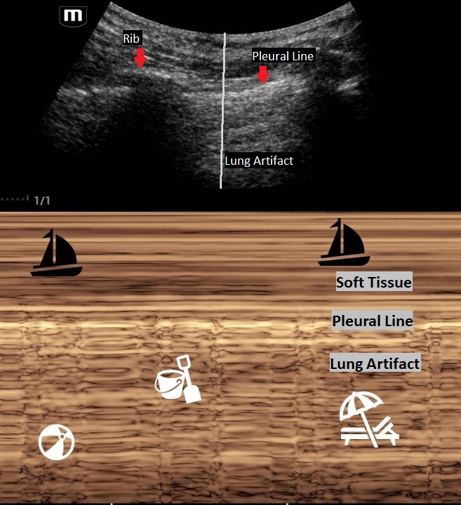
A lung with a pneumothorax will have poor lung sliding. When you image the lung with M-Mode it looks like a classic barcode or "stratosphere sign."
Make sure that you are on "Lung Mode" or decrease the gain to better image the movement of the pleural line. The negative predictive value for lung sliding on ultrasound is 99%. This means that if you see lung sliding you do not have a pneumothorax in that area. However, lung sliding is affected by certain conditions such as blebs, pulmonary fibrosis, pleural adhesions and right mainstem intubation. So, like any other radiology study, clinically correlate!
Thinking about placing a chest tube or have a patient with multiple rib fractures? Take a look at how to perform a Serratus Anteror Plane Block here: https://www.thepocusatlas.com/thoracoabdominal-blocks#Serratus
Did you know that you can use the linear probe with pulse wave (PW) doppler over the femoral artery to look for a pulse during CPR pauses?
Well, the researchers of this article took this skill one step further to evaluate if they could use the femoral artery PW doppler while CPR was in progress to look for signs of a pulse.
The authors found that:
- pulsations due to compressions were organized with uniform pulsations.
- when there was also native cardiac activity, the pulsations were nonuniform and may have an irregular cadence.
Although there were several limitations, Arterial doppler was 100% specific and 50% sensitive in detecting organized cardiac activity during active CPR.
Take Home Point: Take a look at your arterial doppler for signs of organized cardiac activity during a resuscitation.
In this study the researchers looked at patients presenting to the emergency department with high suspicion for ACS and explored if Regional Wall Motion Abnormality (RWMA) evaluation by EPs was associated with occlusion myocardial ischemia (OMI).
FOCUS identified RWMA in 87% of patients with coronary angiography proven OMI. With a sensitivity of 94%, specificity 35%, and overall accuracy of 78%.
The authors concluded that using FOCUS can have good utility when a patient is high risk for OMI and has an equivocal ekg. However, if RWMA is not present, physicians should still continue with work up such as cardiac catheterization.
To evaluate RWMA it is easiest to:
For more information check out this ACEPnow article: https://www.acepnow.com/article/detect-cardiac-regional-wall-motion-abnormalities-point-care-echocardiography/?singlepage=1