Category: Pediatrics
Keywords: meningitis, neck pain, retropharyngeal abscess (PubMed Search)
Posted: 11/16/2012 by Mimi Lu, MD
Click here to contact Mimi Lu, MD
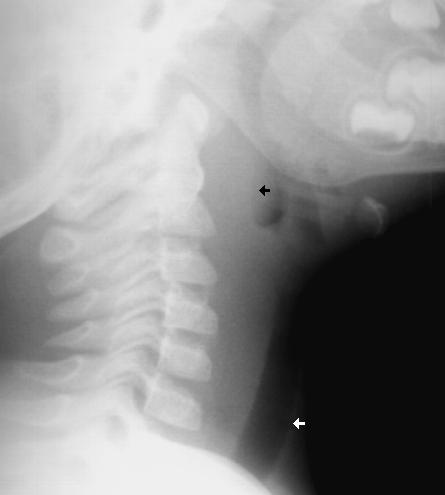
A 1 year old gets sent from their pediatrician’s office for rule out meningitis. They presented with fever for 2 days and neck rigidity. Your LP results are normal. What additional test should you consider?
Answer:
Lateral neck x-ray
http://www.hawaii.edu/medicine/pediatrics/pemxray/v2c20.html

Retropharyngeal abscess (RPA) can commonly present like meningitis. Have a high suspicion in
children who are too young to complain of sore throat or difficulty swallowing.
A recent article in Pediatric Infectious Disease Journal detailed the rising incidence of retropharyngeal abscess, especially in younger patients, which is attributed to community acquired MRSA.
From 2004-2010 there was a 2.8 fold increase in RPA from the previous study period (1993-2003).
Children whose abscess grew MRSA were younger (mean 11 months) than the others (mean 62 months) (P < 0.001) and required longer duration of hospitalization (mean 8.8 days) than the rest (mean 4.5 days) (P = 0.002).
Bottom line: Consider a plain film in the child you are preparing to LP for meningitis.
Reference:
Abdel-Haq, N, Quezada M, Asmar BI. Retropharyngeal abscess in children: the rising incidence
of methicillin-resistant Staphylococcus aureus. Pediatr Infect Dis J 2012; 31: 696–699
Category: Pediatrics
Posted: 11/2/2012 by Jenny Guyther, MD
(Updated: 2/8/2026)
Click here to contact Jenny Guyther, MD
Conventional pediatric nasal cannula can safely deliver up to 4 lpm but are limited by cooling and drying of the airway. This leads to decreased airway patency, nasal mucosal injury, bleeding and possibly increase in coagulase negative staph infections.
HFNC delivers flow up to 40 lpm with 95-100% relative humidity at a controlled temperature. In infants, the initial flow rate is set between 2-4 lpm and can be increased to 8 lpm. Older children and can be started at 10 lpm and increased as high as 40 lpm. Oxygen is also adjustable.
Studies have shown improved comfort, respiratory rate and oxygenation compared to nasal CPAP.
Noninvasive Ventilation Techniques in the Emergency Department: Applications in Pediatric Patients. Pediatric Emergency Medicine Practice. Vol 6 No 6. June 2009.
Spentzas et al. Children with Respiratory Distress Treated with High-Flow Nasal Cannula. Journal of Intensive Care Medicine. Vol 24 No 5. September/October 2009.
Category: Pediatrics
Keywords: croup, laryngomalacia (PubMed Search)
Posted: 10/26/2012 by Mimi Lu, MD
Click here to contact Mimi Lu, MD
Category: Pediatrics
Posted: 10/12/2012 by Rose Chasm, MD
(Updated: 2/8/2026)
Click here to contact Rose Chasm, MD
Glaser N, Barnett P, et al. Risk factors for cerebral edema in children with diabetic ketoacidosis. The Pediatric Emergency Medicine Collaborative Research Committee of the American Academy of Pediatrics. N Engl J Med 2001;344:264.
Category: Pediatrics
Keywords: Vaccines (PubMed Search)
Posted: 10/5/2012 by Jenny Guyther, MD
(Updated: 2/8/2026)
Click here to contact Jenny Guyther, MD
We often ask our pediatric patients if there vaccines are up to date, but what does this mean?
Hepatitis B: birth, 2 and 6 months
Diphtheria/Tetanus and Acellular Pertussis: 2, 4 and 6 months
Pneumococcal vaccine: 2, 4 and 6 months
Haemophilus influenzae B : 2, 4 and 6 months
Polio: 2, 4 and 6 months
Rotavirus: 2 and 4 months or 2, 4 and 6 months depending on the brand.
Influenza: 6 months and older
Children less than 8 years old should receive 2 doses of flu vaccine at least 4 weeks apart during the first flu season that they are immunized. Children older than 2 years are eligible for the nasal vaccine if they do not have asthma, wheezing in the past 12 months or other medical conditions that predispose them to flu complications.
To see the full vaccine schedule including exact time frames between doses and catch up schedules, see: http://www.cdc.gov/vaccines/
Category: Pediatrics
Keywords: dysrhythmia, arrhythmia (PubMed Search)
Posted: 9/28/2012 by Mimi Lu, MD
Click here to contact Mimi Lu, MD
The incidence of pediatric syncope is common with 15%-25% of children and adolescents experiencing at least one episode of syncope before adulthood. Incidence peaks between the ages of 15 and 19 years for both sexes.
Although most causes of pediatric syncope are benign, an appropriate evaluation must be performed to exclude rare life-threatening disorders. In contrast to adults, vasodepressor syncope (also known as vasovagal) is the most frequent cause of pediatric syncope (61%–80%). Cardiac disorders only represent 2% to 6% of pediatric cases but account for 85% of sudden death in children and adolescent athletes. 17% of young athletes with sudden death have a history of syncope.
Key features on history and physical examination for identifying high-risk patients include exercise-related symptoms, a family history of sudden death, a history of cardiac disease, an abnormal cardiac examination, or an abnormal ECG.
Category: Pediatrics
Keywords: premedication, RSI, ventilator, high flow nasal cannula (PubMed Search)
Posted: 9/21/2012 by Mimi Lu, MD
Click here to contact Mimi Lu, MD
Category: Pediatrics
Posted: 9/15/2012 by Rose Chasm, MD
(Updated: 2/8/2026)
Click here to contact Rose Chasm, MD
Category: Pediatrics
Keywords: cervical spine, trauma, pediatrics (PubMed Search)
Posted: 9/7/2012 by Lauren Rice, MD
Click here to contact Lauren Rice, MD
Ligamentous laxity is increased in children and ligamentous injury is more common than fractures.
If fractures occur, they are more likely to be in the upper cervical spine in infants and the lower cervical spine in older children.
Pseudosubluxation: physiologic subluxation between C2-3 and C3-4 may exist until age 16 years
Screening Assessment/Clearance for Verbal Children
-Midline C-spine tenderness?
-Pain with active motion?
-Altered level of alertness?
-Evidence of intoxication?
-Focal neurological deficit?
-Distracting painful injury?
-High impact injury?
Screening Assessment/Clearance for Pre-Verbal Children
-Neurological assessment of basic reflexes
-Response to painful stimuli
-Equal movements of all extremities
-Response to sound (eye tracking)
-Extremity strength and resistance
-Palpate posterior C-spine (observe for facial grimace)
-Feel for step-offs, deformities
-Verify full range of motion of neck (may need to be creative)
-Repeat neurological assessment
If concern arises on screening assessment, keep child in hard cervical collar and image (may start with x-ray and progress to CT if still concerned and x-rays negative).
If imaging negative, but persistent suspicion based on neurological deficits consider SCIWORA (Spinal Cord Injury WithOut Radiographic Abnormality) which exists in up to 50% of children with cervical cord injury, and may require MRI to further identify injury.
Category: Pediatrics
Keywords: septic shock, fluid resuscitation, PALS (PubMed Search)
Posted: 8/31/2012 by Mimi Lu, MD
Click here to contact Mimi Lu, MD
Category: Pediatrics
Posted: 8/24/2012 by Mimi Lu, MD
Click here to contact Mimi Lu, MD
Types:
- Uniphasic anaphylaxis: occuring immediately after exposure to allergen, resolves over minutes to hours and does not recur
- Biphasic anaphylaxis: occuring after apparent resolution of symptoms typically 8 hours after the first reaction. Occur in up to 23% of adults and up to 11% of children with anaphylaxis
Treatment:
1. First line: IM epinephrine 1:1000 solution
- vasoconstrictor effects on hypotension and peripheral vasodilation; bronchodilator effects on upper respiratory obstruction
- NO absolute contraindication for use in anaphylaxis
- Dosage: Adult: 0.3 - 0.5mg; Peds: 0.01mg/kg (max 0.3mg)
- can be repeated every 5-15 minutes
2. Adjunctive therapy:
- H1 Blocker: diphenhydramine 1-2mg/kg up to 50mg IV
- H2 Blocker: ranitidine 1-2mg/kg
- Corticosteroid: 1-2 mg/kg for prevention of biphasic reactions
- Bronchodilator: Albuterol for bronchospasm
- Glucagon: for refractory hypotension or if patient is on beta blocker
- Dosage: Adult: 1-5 mg; Peds 20-30microgm/kg
- Dose may be repeated or followed by infusion of 5-15 mg/min
- place patient in recumbent position if tolerated with lower extremities elevated
- supplemental O2
- IV fluids for hypotension
Fatalities: typically seen with peanut or treenut ingestions from cardiopulmonary arrest. Associated with delayed or inappropriate epinephrine dosing
Disposition:
- Mild reaction with symptom resolution: observe for 4-6 hrs (ACEP, AAP)
- Recurrent symptoms or incomplete resolution: admit
Reference:
1. World Allergy Organization Guidelines for the Assessment and Management of Anaphylaxis, Feb 2011
2. Guidelines for the Diagnosis and Management of Food Allergy in the United States: Report of the NIAID-Sponsored Expert Panel Oct 2010
Category: Pediatrics
Keywords: vaccination, whooping cough (PubMed Search)
Posted: 8/17/2012 by Mimi Lu, MD
Click here to contact Mimi Lu, MD
If you have a patient who meets (or has had close exposure to someone meeting) the clinical case definition of pertussis (a cough lasting at least 2 weeks with one of the following: paroxysms of coughing, inspiratory “whoop,” or post-tussive vomiting) here are some important points to keep in mind:
Vaccination
Testing
Treatment
References:
Altunaiji SM, Kukuruzovic RH, Curtis NC, Massie J. Antibiotics for whooping cough (pertussis). Cochrane Database of Systematic Reviews 2007, Issue 3. Art. No.: CD004404. DOI: 10.1002/14651858.CD004404.pub3
http://www.cdc.gov/vaccines/pubs/surv-manual/chpt10-pertussis.html
Category: Pediatrics
Posted: 8/10/2012 by Rose Chasm, MD
Click here to contact Rose Chasm, MD
Category: Pediatrics
Posted: 8/3/2012 by Lauren Rice, MD
(Updated: 2/8/2026)
Click here to contact Lauren Rice, MD
Henoch-Schonlein Purpura (aka. Anaphylactoid purpura) is a small vessel vasculitis.
Background:
Clinical Features:
Etiology:
Diagnosis:
Treatment:
Category: Pediatrics
Keywords: hemolysis, bilirubin, kernicterus, jaundice (PubMed Search)
Posted: 7/27/2012 by Mimi Lu, MD
Click here to contact Mimi Lu, MD
Bonus pearl: Types of Jaundice by Age
- < 24 hrs: hemolyis, TORCH, bruising from birth trauma (ie- cephalohematoma), acquired infection
- Day 2-3: Physiologic
- Day 3-7: infection, congenital diseases, TORCH
- >1 week: Breast Milk Jaundice, breast feeding jaundice, drug hemolysis, hypothyroidism, biliary atresia, hepatitis, red cell membrane disorders (SS, HS, G6PD deficiency)
Category: Pediatrics
Keywords: leukemia, back pain, cancer (PubMed Search)
Posted: 6/29/2012 by Mimi Lu, MD
(Updated: 7/20/2012)
Click here to contact Mimi Lu, MD
Category: Pediatrics
Posted: 7/13/2012 by Rose Chasm, MD
(Updated: 2/8/2026)
Click here to contact Rose Chasm, MD
NMS Pediatrics, 4th edition
Category: Pediatrics
Posted: 6/29/2012 by Rose Chasm, MD
(Updated: 2/8/2026)
Click here to contact Rose Chasm, MD
Submitted by Dr. Lauren Rice
The summertime can be full of lots of fun activities (beach, fireworks, cookouts, and campfires) that can put children at risk of burns.
Burn depth classification:
1. Superficial (first-degree): red and blanching with minor pain, resolves in 5-7 days
2. Partial thickness (second-degree): red and wet with blisters, very painful, resolves in 2-5 weeks
Treatment: clean with soap and water twice daily, and apply silvadene wrap with gauze, kerlex
3. Full thickness (third-degree): dry and leathery without pain, no resolution after 5-6 weeks, may require graft
Treatment: wound debridement and dressings as above
Parkland formula: 4ml/kg/%TBSA in 1st 24 hours with 50% of total volume in 1st 8 hours
Calculate burn surface area:
-SAGE: free computerized burn diagram available at www.sagediagram.com
-Rule of Nines > 14 years old
-Rule of Palm <10 years old
Burn Center Referral
-Extent: partial thickness of >30% TBSA or full thickness of >10-20%
-Site: hands, feet, face, perineum, major joints
-Type: electrical, chemical, inhalation
1. Cross, J.T. and Hannaman, R.A. MedStudy Pediatrics Board Review Core Curriculum, 5th edition, p. 3-11, 3-12.
2. Children’s National Medical Center, Department of Trauma and Burn Surgery. Trauma Cheat Sheet.
Category: Pediatrics
Posted: 6/23/2012 by Mimi Lu, MD
(Updated: 6/29/2012)
Click here to contact Mimi Lu, MD
Pathology at the umbilicus can manifest as inflammation, drainage, a palpable mass, or herniation.
Omphalitis - A cellulitis of the umbilicus. Mild cases often respond to local application of alcohol to clean the area, but due to the possibility of rapid progression and abdominal wall necrotizing fasciitis, admission for observation and IV antibiotics is usually warranted. Cover staph, strep, and GNRs.
Umbilical granuloma - As the umbilical ring closes and the cord sloughs off, granulation tissue formation is a normal part of umbilical epithelialization. There is sometimes an overgrowth of granulation tissue which can be treated once or twice with silver nitrate. Should the tissue not regress after a 1-2 treatments, the patient should be referred to pediatric surgery for excision and evaluation of other pathology (urachal or vitelline remnants).
Umbilical fistula - This is a patent vitelline duct and is characterized by persistent drainage that is bilious or purulent. A fistulogram using a small catheter and radio opaque dye can sometimes be helpful in determining the source of drainage (dye should be seen in the small bowel).
Umbilical polyp - Often confused with an umbilical granuloma with its glistening cherry red appearance, this is actually a vitelline duct remnant and contains small bowel mucosa. It does not regress with silver nitrate.
Vesicoumbilical fistula/sinus - The urachal versions of the umbilical fistula. This are a failure of complete closure of the urachus, resulting in persistent drainage of urine from the umbilicus, and infection (including recurrent UTIs). A fistulogram can be helpful for diagnosis.
Category: Pediatrics
Keywords: abdominal pain, vomiting, bloody stool, altered mental status, lethargy (PubMed Search)
Posted: 6/22/2012 by Mimi Lu, MD
Click here to contact Mimi Lu, MD
Intussusception is the telescoping or prolapse of one portion of the bowel into an immediately adjacent segment.
