Category: Visual Diagnosis
Posted: 10/25/2016 by Tu Carol Nguyen, DO
(Updated: 10/26/2016)
Click here to contact Tu Carol Nguyen, DO
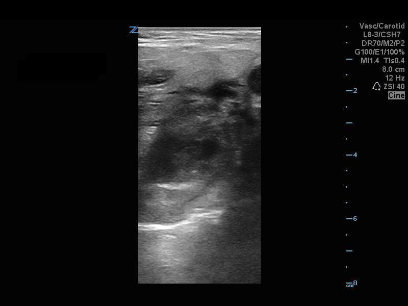
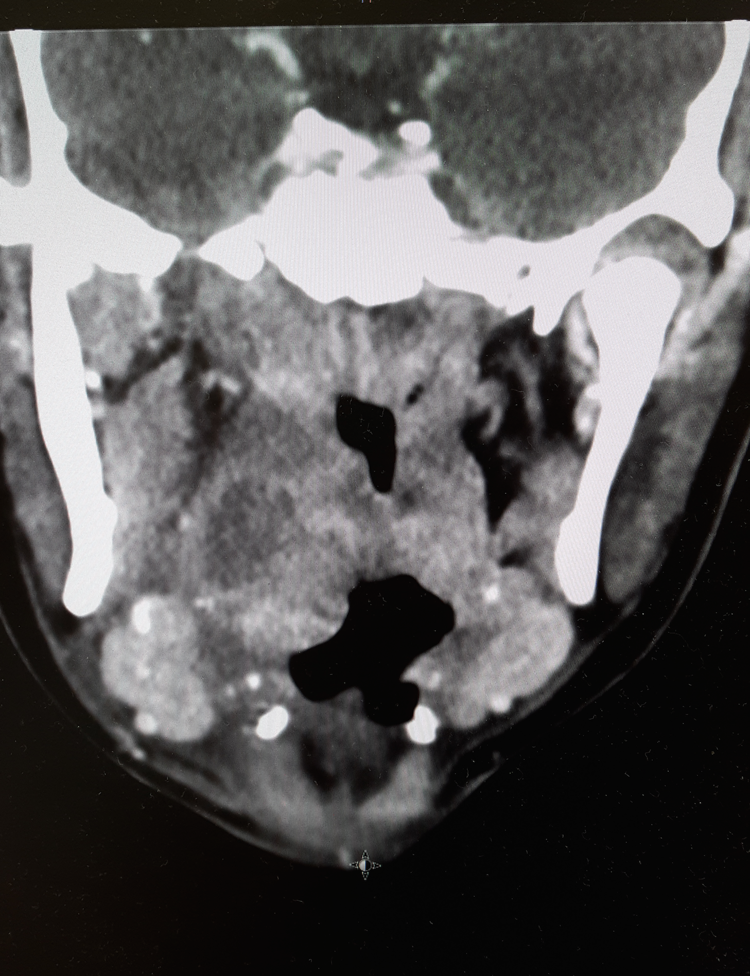
20 year-old female presents with sore throat, right throat fullness, difficulty speaking for 2-3 days. A bedside ultrasound and subsequent CT was obtained as seen below. What's the diagnosis?
Peritonsillar Abscess
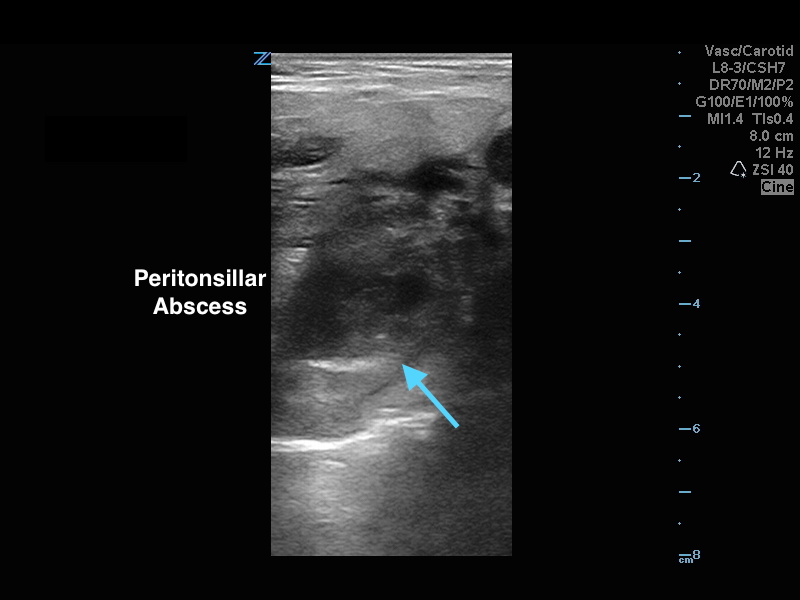
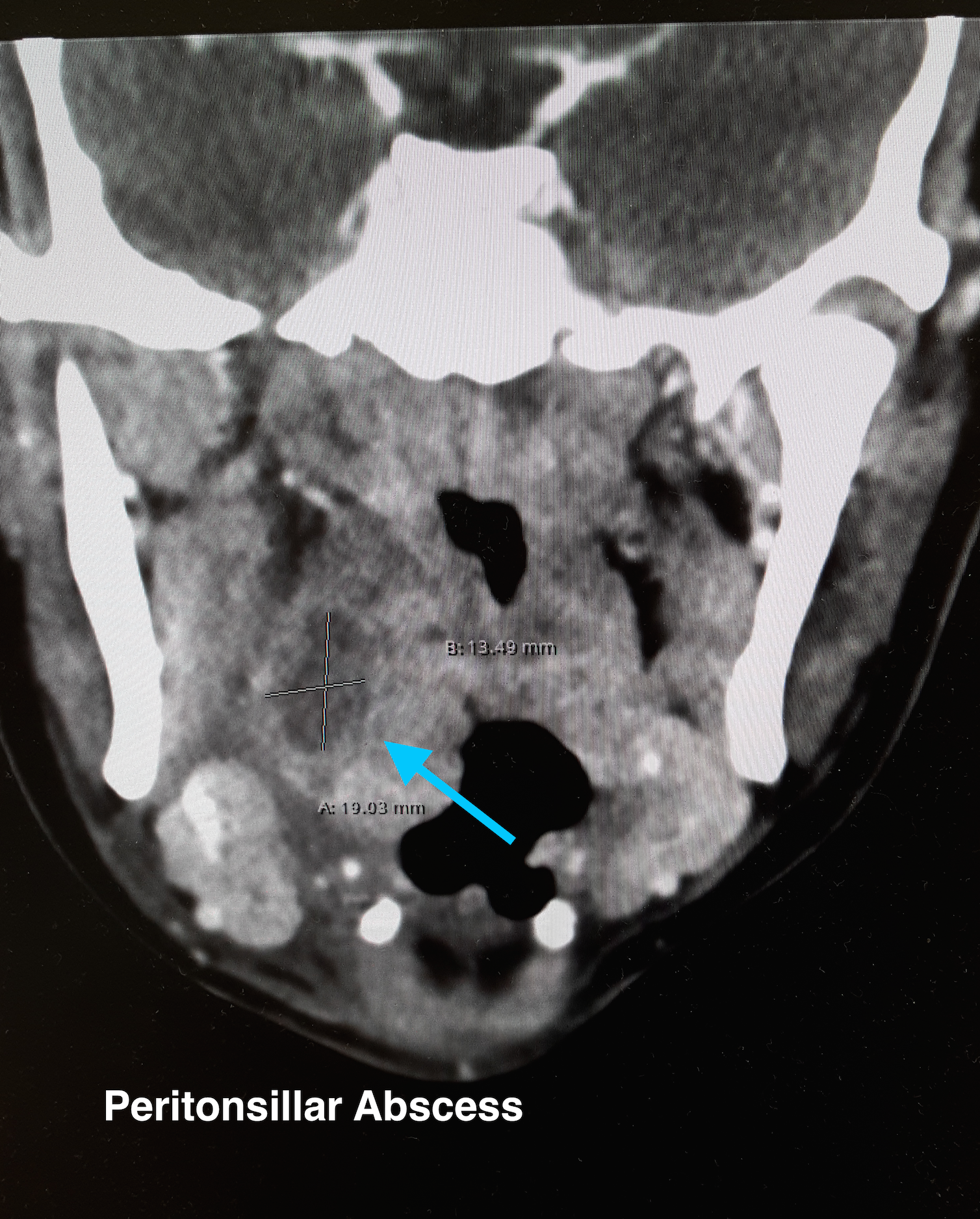
The ultrasound image is a transcutaneous approach with a linear transducer that is placed at the angle of the mandible of the affected side. This is an alternative approach to an intra-oral ultrasound with the endocavitary transducer if the patient has trismus.
Take Home Points:
How to do an intra-oral US-guided needle aspiration of PTA, check out:
http://www.ultrasoundpodcast.com/2012/01/episode-21-full-peritonsillar-abscess-podcast/
For a brief video on how to perform a transcutaneous US for PTA:
https://www.youtube.com/watch?v=JkIYOhKCweI&t=28s
Halm BM, Ng C, Larrabee YC. Diagnosis of a Peritonsillar Abscess by Transcutaneous Point-of-Care Ultrasound in the Pediatric Emergency Department. Pediatr Emerg Care. 2016;32(7):489-92.
Rehrer M, Mantuani D, Nagdev A. Identification of peritonsillar abscess by transcutaneous cervical ultrasound. Am J Emerg Med. 2013;31(1):267.e1-3.
Category: Visual Diagnosis
Posted: 10/10/2016 by Tu Carol Nguyen, DO
Click here to contact Tu Carol Nguyen, DO
57 year-old female with history of bilateral lung transplants presents with fever, and 2 days of a painful, red, bumpy rash over the left labia and left buttock, but also notes a small tender area on the plantar surface of the left foot.
Below is a figure depicting the location of the rash, as well as a photo of her foot.
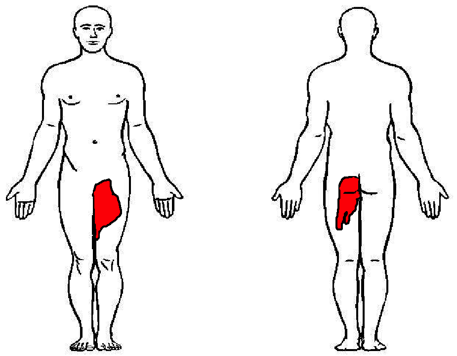
This is Herpes Zoster (Shingles).
Presentation:
Treatment:
Dworkin RH, Johnson RW, Breuer J, et al. Recommendations for the management of herpes zoster. Clin Infect Dis. 2007;44 Suppl 1:S1.
Category: Visual Diagnosis
Posted: 10/3/2016 by Hussain Alhashem, MBBS
Click here to contact Hussain Alhashem, MBBS
A 41 year old female presenting with intermittent RUQ abdominal pain for 1 week. An ultrasound of the right upper quadrant was performed. What is the diagnosis ?
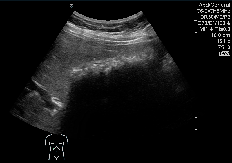
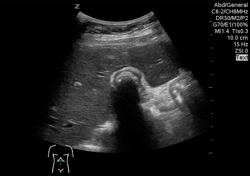
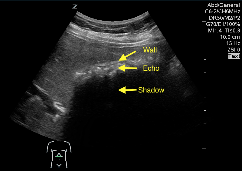
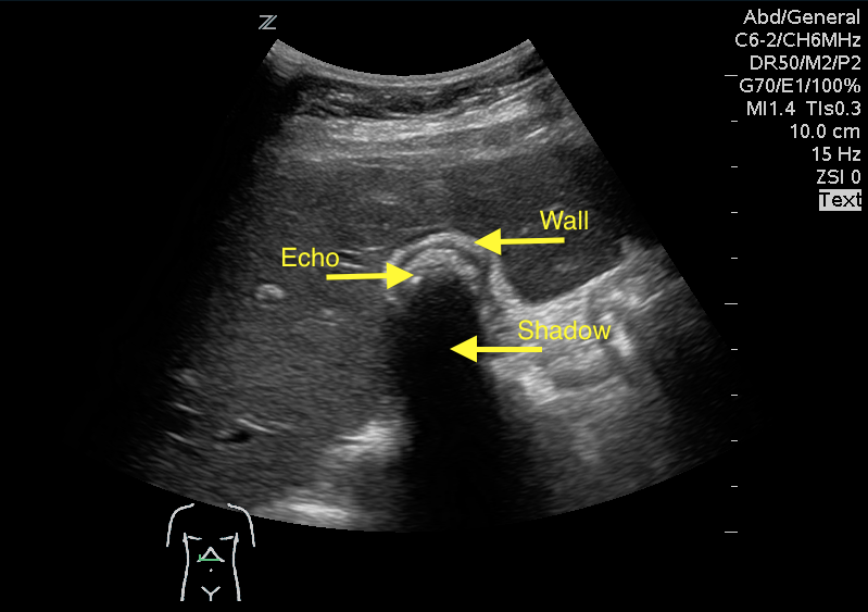
Answer: WES sign
WES sign stands for Wall Echo Shadow sign. It is a triad of:
1- Thick echogenic gall bladder wall (W)
2- Echoes filling the gallbladder (E)
3- A posterior acoustic shadow (S)
Rybicki, F. J. (2000). The WES Sign 1. Radiology, 214(3), 881-882.
Category: Visual Diagnosis
Posted: 9/14/2016 by Tu Carol Nguyen, DO
(Updated: 9/26/2016)
Click here to contact Tu Carol Nguyen, DO
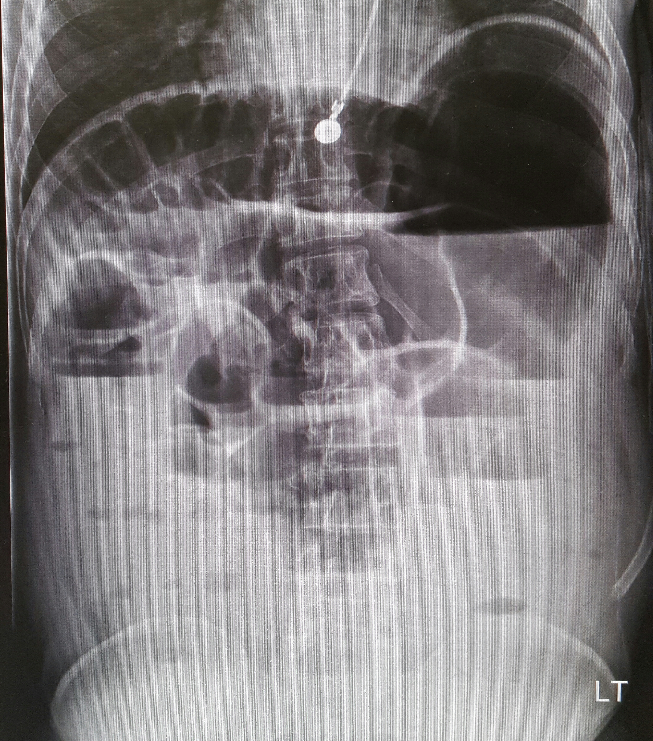
22-year-old male with history of autism, mental retardation who is non-verbal presents with abdominal pain and vomiting for one day. Patient was found clutching his abdomen and moaning. What's the diagnosis?
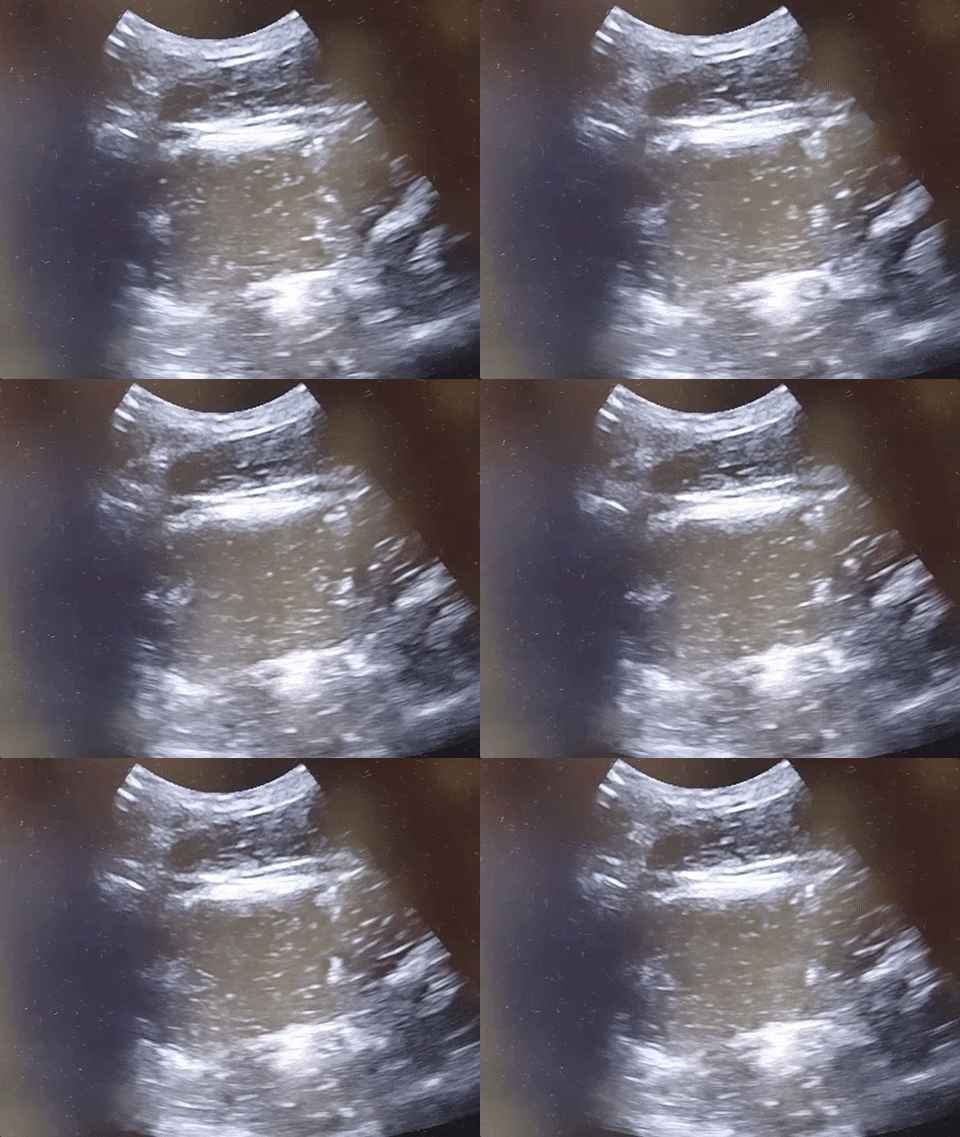
Small Bowel Obstruction
See the corresponding upright abdominal x-ray, showing dilated bowel with air fluid levels.
Kameda T, Taniguchi N. Overview of point-of-care abdominal ultrasound in emergency and critical care. J Intensive Care. 2016 Aug 15;4:53. doi: 10.1186/s40560-016-0175-y. eCollection 2016. Review.
Unl er EE, Yava i O, Ero lu O, Yilmaz C, Akarca FK. Ultrasonography by emergency medicine and radiology residents for the diagnosis of small bowel obstruction. Eur J Emerg Med. 2010 Oct; 17(5):260-4.
Category: Visual Diagnosis
Posted: 9/19/2016 by Hussain Alhashem, MBBS
Click here to contact Hussain Alhashem, MBBS
A 67 year old female with history of CVA, presented from a nursing home with RUQ abdominal pain and inablitiy to tolerate PO for 3 days. A CT scan of her abdomen was obtained. What is the diagnosis ?
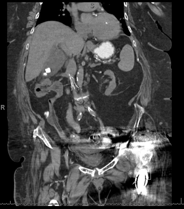
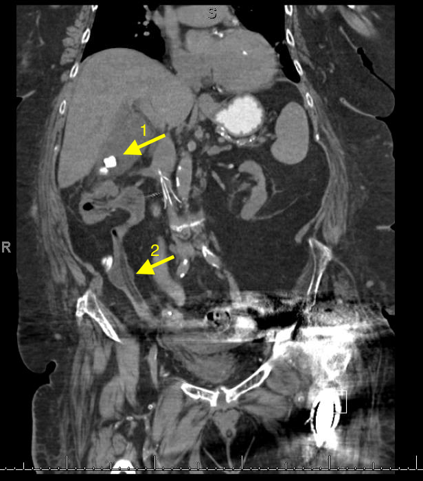
1- Cholecystitis
Ultrasound remains the best modality to test for cholecystitis. However, CT scans can still be obtained for non-classic presentations. The negative predictive value of CT is still relatively high. CT has a negative predictive value of 89%, compared to 97% to that of ultrasound. The absence of cholecysitis on CT will help the argument against the diagnosis, but if the suspicion is high an ultrasound study should still be obtained.
Things to look for on an abdominal CT that are suggestive of cholecysitis:
Gallbladder distension ( >5 cm width, >8 cm length).
Wall thickening ( >4mm thickness).
Pericholecystic fat stranding.
Presence of gallstones.
2- An inflated foley catheter in the ureter!
Ureteric insertion of foley catheters is a very rare complication of foley catheterization. There are no clear predisposing factors to this complication. However, it is thought that the presence of an underlying anatomical deformity (e.g. abnormal ureteric insertion site) might put the patient at a higher risk for it. Inflating a balloon in the ureter might result in severe ureteric injury. A suggested method to prevent this kind of injury is to perform bladder aspiration to insure balloon positioning prior to inflation.
References
1- Shakespear, Jonathan S., Akram M. Shaaban, and Maryam Rezvani. "CT findings of acute cholecystitis and its complications." American Journal of Roentgenology 194.6 (2010): 1523-1529.
2- Kim, Myung Ki, and Kwangsung Park. "Unusual complication of urethral catheterization: a case report." Journal of Korean medical science 23.1 (2008): 161.
Category: Visual Diagnosis
Posted: 9/8/2016 by Tu Carol Nguyen, DO
(Updated: 9/12/2016)
Click here to contact Tu Carol Nguyen, DO
A 25-year-old male was brought in by EMS with a stab wound to the chest. What's the diagnosis?
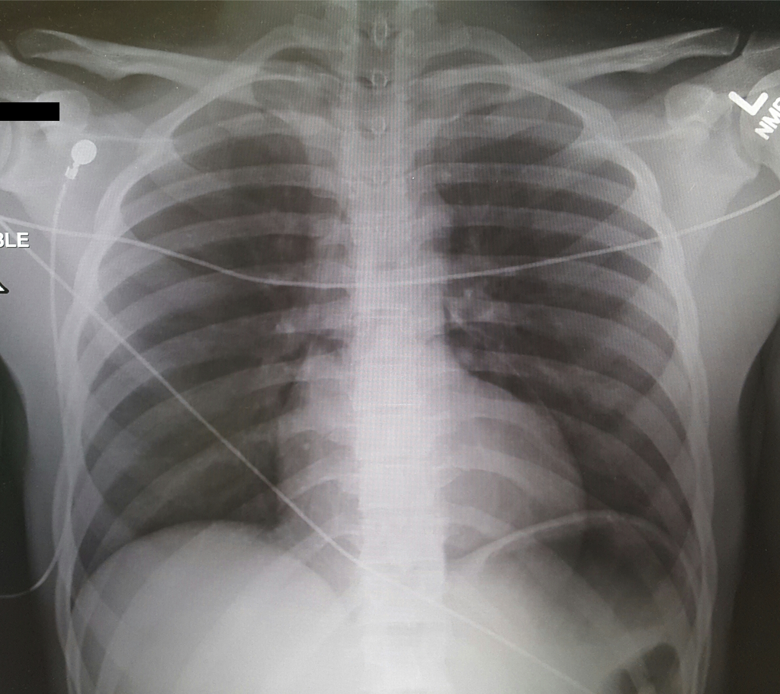
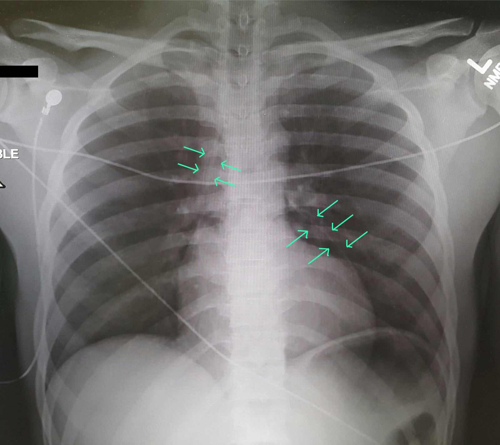
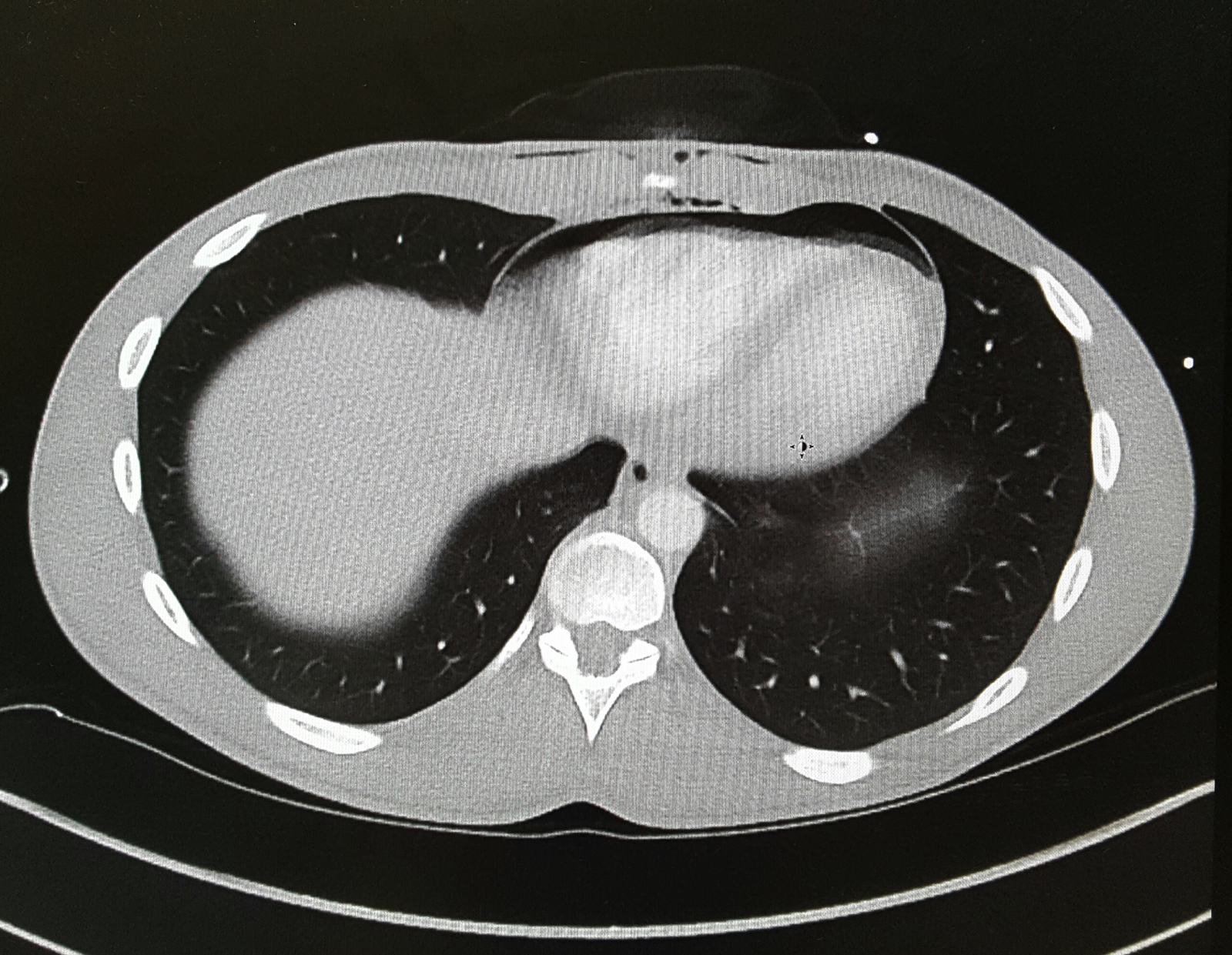
Differential Diagnosis: Bronchial injury, Diaphragm injury, Hemothorax, Tension Pneumothorax, Aortic Transection, Esophageal injury, Pneumomediastinum
Evaluation: Ultrasound (FAST Exam), CXR, CTA in stable patients, ECG, troponin.
Management: Penetrating cardiac trauma require emergent thoracotomy, pericardial window.
Clancy K, Velopulos C, Bilaniuk JW, et al. Screening for blunt cardiac injury: an Eastern Association for the Surgery of Trauma practice management guideline. J Trauma Acute Care Surg. 2012;73(5 Suppl 4):S301-6.
El-menyar A, Al thani H, Zarour A, Latifi R. Understanding traumatic blunt cardiac injury. Ann Card Anaesth. 2012;15(4):287-95.
Tintinalli's 7th Edition. Emergency Medicine Manual. Chapter 164: Cardiothoracic Trauma.
Category: Visual Diagnosis
Posted: 2/29/2016 by Haney Mallemat, MD
Click here to contact Haney Mallemat, MD
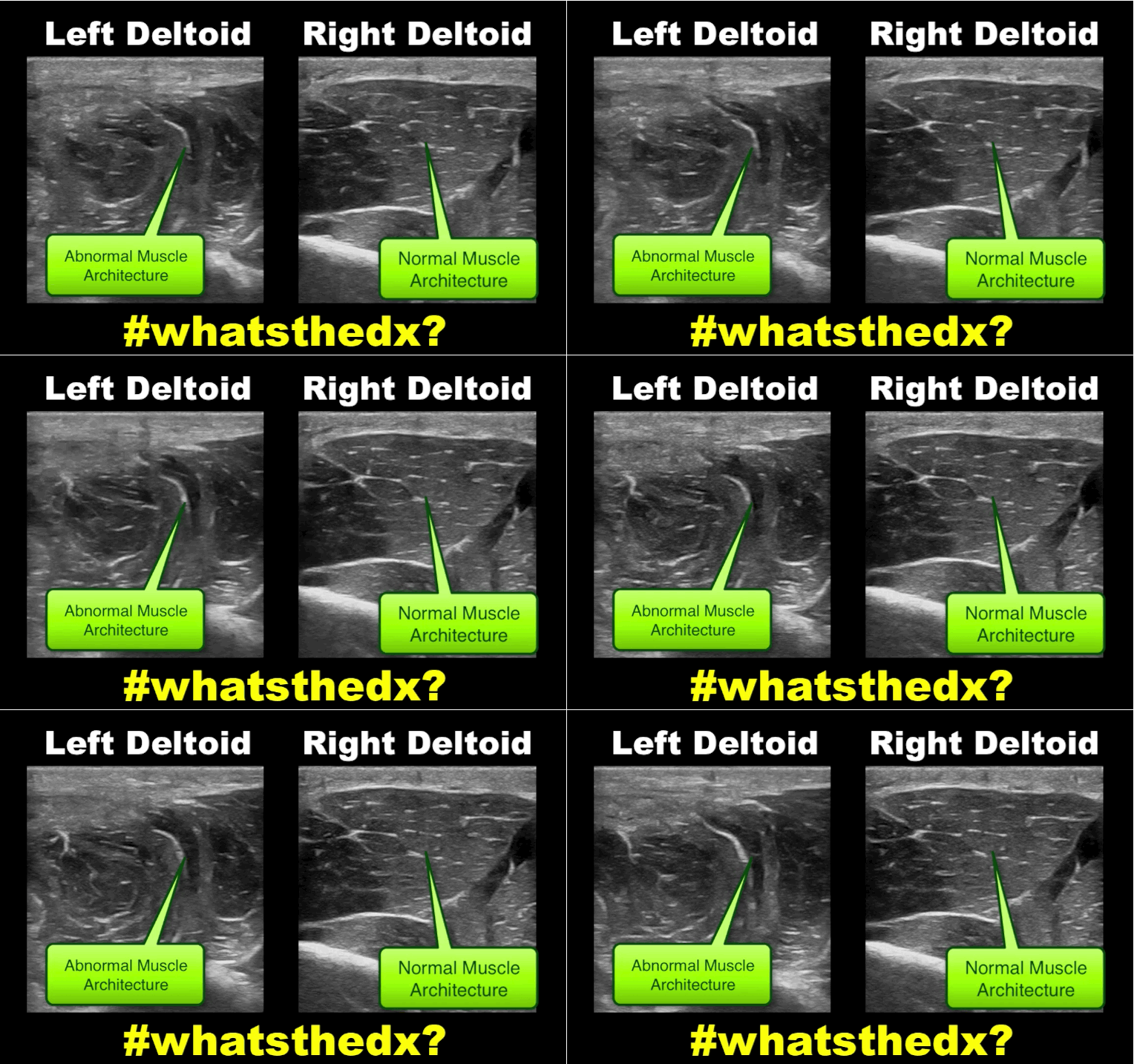
19 year-old male complaining of left arm pain one week after injecting anabolic steroids into his shoulder. What's the diagnosis?
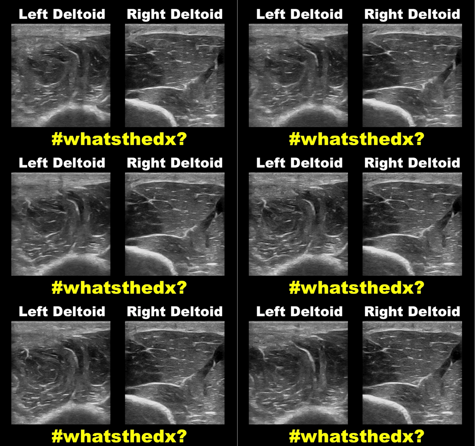
Myositis of the deltoid muscle
Follow me on Twitter (@criticalcarenow)
Category: Visual Diagnosis
Posted: 1/18/2016 by Haney Mallemat, MD
Click here to contact Haney Mallemat, MD
23 year-old female presents complaining of progressive right lower quadrant pain after doing "vigorous" exercise. CT abdomen/pelvis below. What’s the diagnosis? (Hint: it’s not appendicitis)
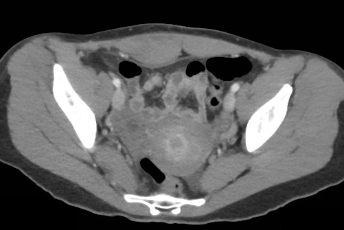
Rectus sheath hematoma
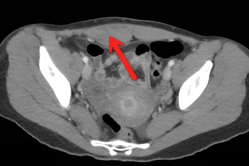
Rectus Sheath Hematoma (RSH)
Rectus muscle tear causing damage to the superior or inferior epigastric arteries with subsequent bleeding into the rectus sheath; uncommon cause of abdominal pain but mimics almost any abdominal condition.
Diagnose with CT, but try using ultrasound (thanks Dr. Joseph Minardi)
May occur spontaneously, but suspect with the following risk factors:
Typically a self-limiting condition, but hypovolemic shock may result from significant hematoma expansion.
Hemodynamically stable (non-expanding hematoma): conservative treatment (rest, analgesia, and ice)
Hemodynamically unstable (expanding hematoma): treat with fluid resuscitation, reversal of coagulopathy, and transfusion of blood products.
Follow me on Twitter (@criticalcarenow)
Category: Visual Diagnosis
Posted: 1/11/2016 by Haney Mallemat, MD
(Updated: 3/10/2016)
Click here to contact Haney Mallemat, MD
What’s the name of this CT finding and name two potential causes?

Pneumobilia (air in the biliary tree). Be careful, this must be distinguished from portal venous gas.
Diagnoses to consider when pneumobilia is present:
Category: Visual Diagnosis
Posted: 12/28/2015 by Haney Mallemat, MD
Click here to contact Haney Mallemat, MD
79 year-old male with headaches, ataxia, falls, and difficulty urinating. What's the diagnosis?
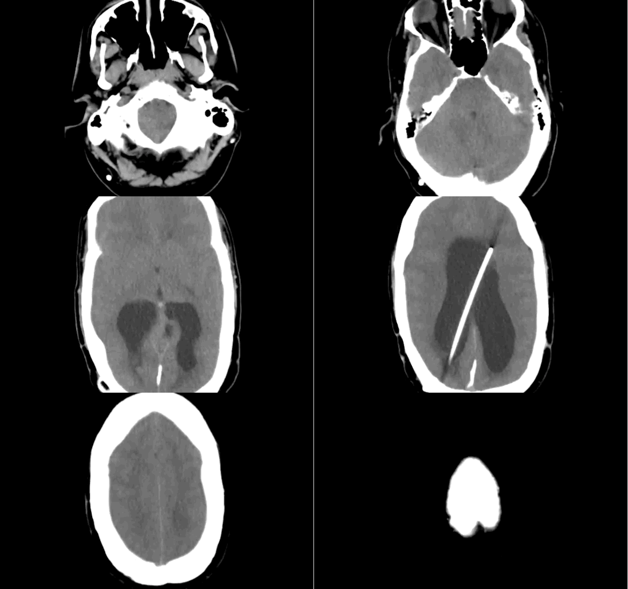
Diagnosis: Ventriculomegaly secondary to Normal Pressure Hydrocephalus
An approach to ventriculomegaly
Ventriculomegaly is due to cerebral atrophy (e.g., Parkinson disease) or increased cerebrospinal fluid (CSF) within the ventricles. Increased CSF is due to:
Congenital causes of ventriculomegaly:
Acquired causes of ventriculomegaly:
Follow me on Twitter (@criticalcarenow)
Category: Visual Diagnosis
Posted: 12/15/2015 by Haney Mallemat, MD
Click here to contact Haney Mallemat, MD
A patient arrives in acute respiratory distress with left sided chest pain. Ultrasound of the left anterior chest is shown; what's the diagnosis and name one false positive?
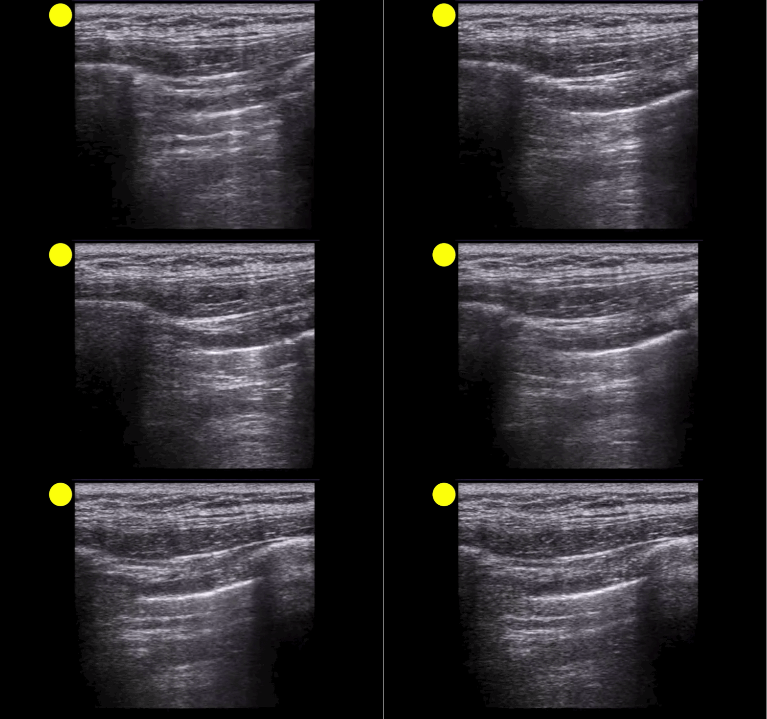
Lung point indicating pneumothorax (PTX)....see below for the false positives
What's the (Lung) Point
Follow me on Twitter (@criticalcarenow)
Category: Visual Diagnosis
Posted: 12/14/2015 by Haney Mallemat, MD
Click here to contact Haney Mallemat, MD
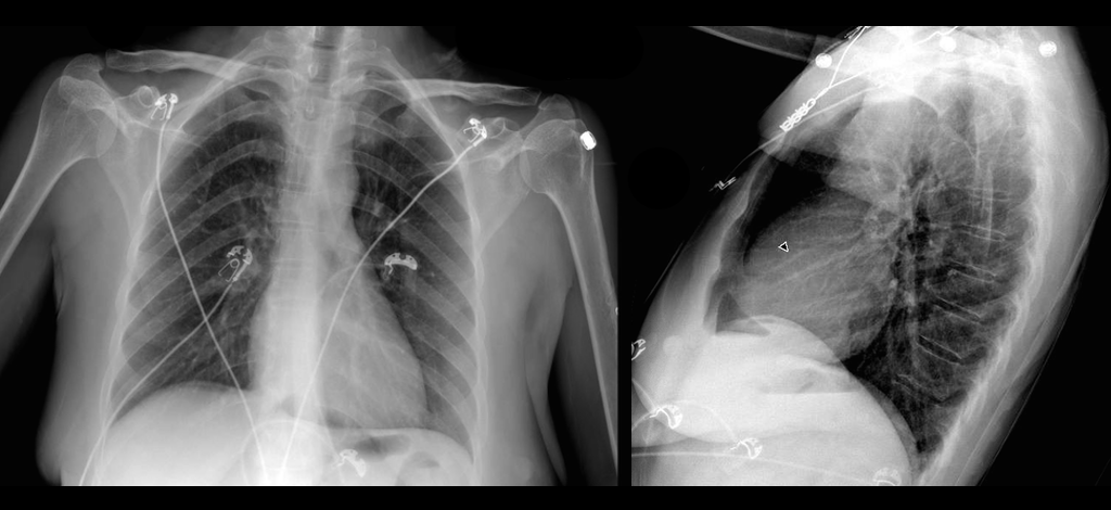
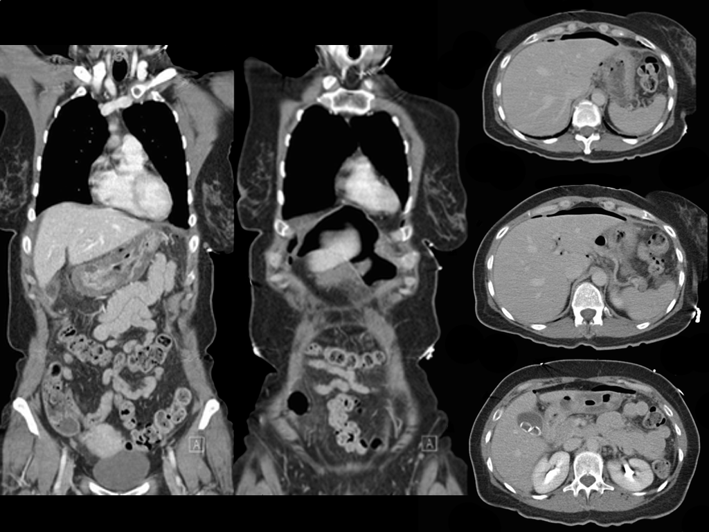
Ruptured gastric ulcer with pneumoperitoneum (CT scan below)
Follow me on Twitter (@criticalcarenow)
Category: Visual Diagnosis
Posted: 12/7/2015 by Haney Mallemat, MD
(Updated: 12/8/2015)
Click here to contact Haney Mallemat, MD
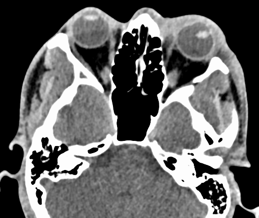
Lens disclocation
Follow me on Twitter (@criticalcarenow)
Category: Visual Diagnosis
Posted: 11/30/2015 by Haney Mallemat, MD
Click here to contact Haney Mallemat, MD
Patient presents with right elbow pain after a fall. What's the diagnosis and what other injury should you look for?
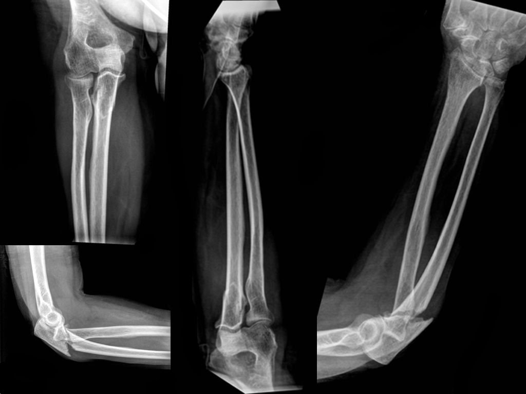
Displaced fracture of the proximal ulna.
Pearls
Follow me on Twitter (@criticalcarenow)
Category: Visual Diagnosis
Posted: 11/23/2015 by Haney Mallemat, MD
(Updated: 12/5/2015)
Click here to contact Haney Mallemat, MD
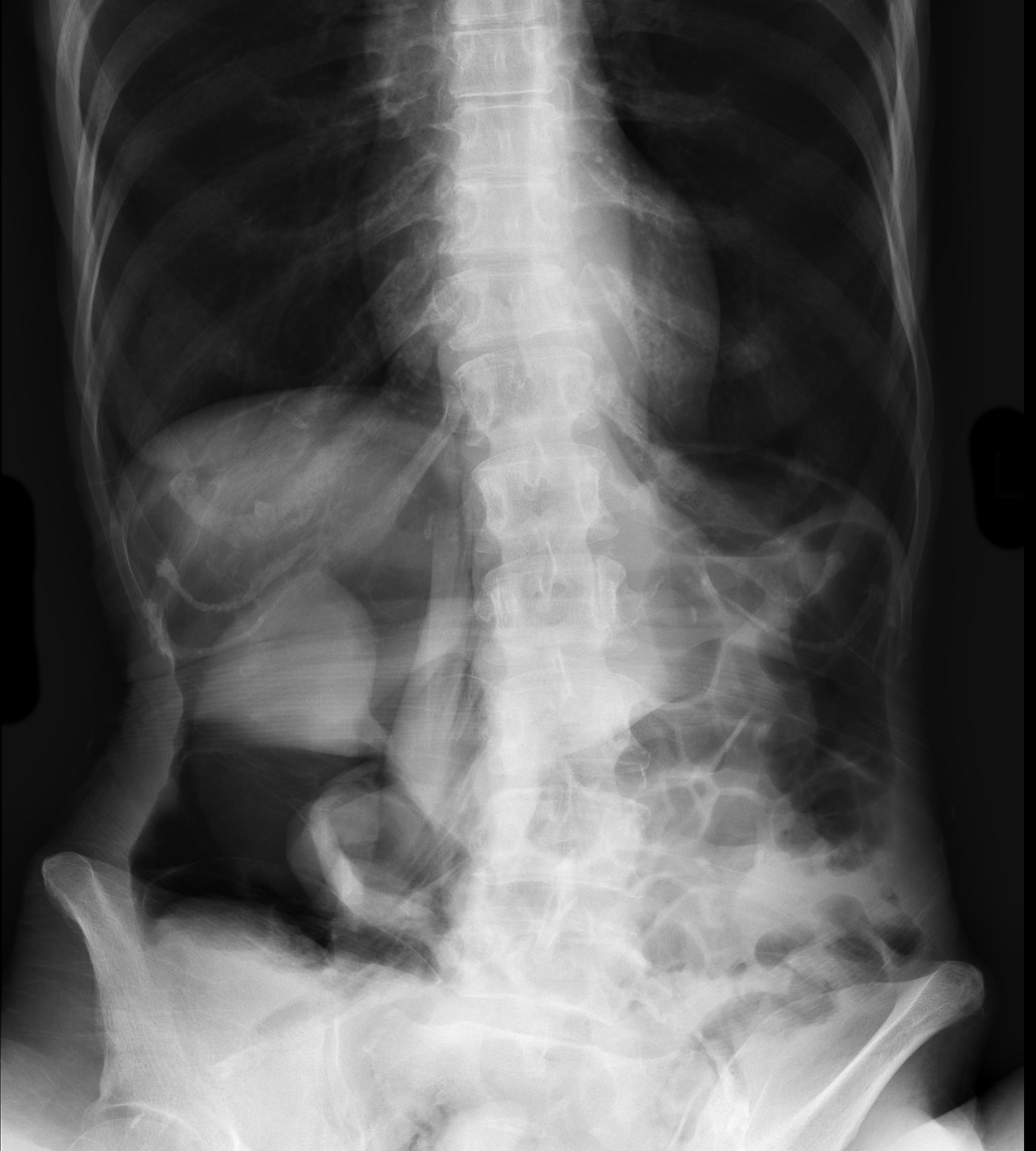
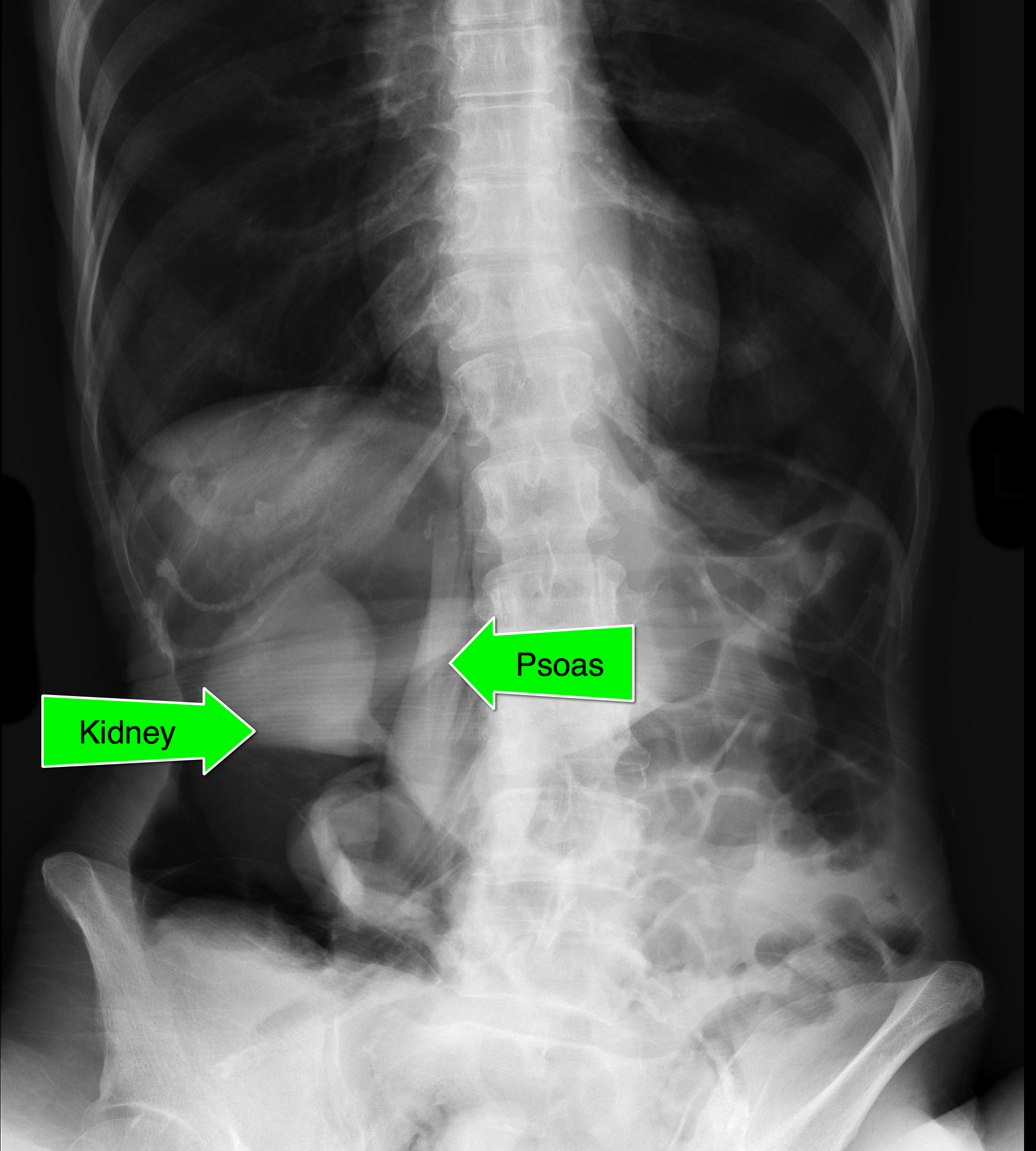
An elderly patient presents with a history of weight loss and chronic constipation. The abdominal Xray is shown below. What's the diagnosis?
This one is tricky so here's a hint: why is the right kidney and psoas muscle so well defined?
Follow me on Twitter (@criticalcarenow)
Category: Visual Diagnosis
Posted: 11/1/2015 by Haney Mallemat, MD
Click here to contact Haney Mallemat, MD
Patient complains of facial and neck swelling, what's the diagnosis?

Subcutaneous emphysema
Follow me on Twitter (@criticalcarenow)
Category: Visual Diagnosis
Posted: 10/19/2015 by Haney Mallemat, MD
Click here to contact Haney Mallemat, MD
8 year-old female presents with nausea, vomiting, double-vision and inability to move her left eye upwards after being kicked in the face at school. What's the diagnosis?
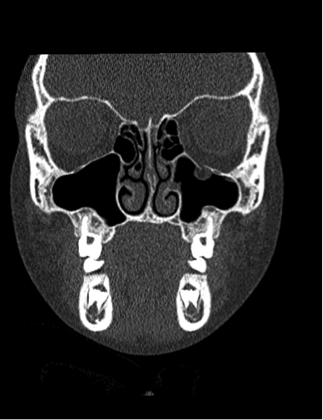
Orbital floor fracture with entrapment of the inferior rectus muscle.
Follow me on Twitter (@criticalcarenow)
Category: Visual Diagnosis
Posted: 10/12/2015 by Haney Mallemat, MD
Click here to contact Haney Mallemat, MD
5 year-old boy who presents with sudden onset hoarse voice, and drooling without a fever.
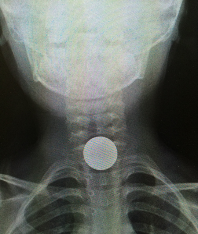
Coin lodged in the esophagus
Coin ingestions
Follow me on Twitter (@criticalcarenow)
Category: Visual Diagnosis
Posted: 10/5/2015 by Haney Mallemat, MD
(Updated: 10/7/2015)
Click here to contact Haney Mallemat, MD
Patient presents after being started on an antibiotic for cellutlitis of lower extremity. What's the diagnosis and what are some other etiologic agents (name 3)
Erythema Multiforme (minor)
Follow me on Twitter (@criticalcarenow)
Category: Visual Diagnosis
Posted: 9/28/2015 by Haney Mallemat, MD
Click here to contact Haney Mallemat, MD
26 year-old male presents with a swollen 4th digit and pain during extension, what’s the diagnosis?
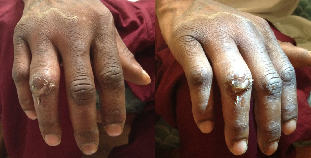
Answer: Infectious Flexor Tenosynovitis
Infectious Flexor Tenosynovitis
Follow me on Twitter (@criticalcarenow)
