Category: Ophthamology
Keywords: POCUS, Ocular, Posterior Chamber (PubMed Search)
Posted: 8/7/2023 by Alexis Salerno Rubeling, MD
Click here to contact Alexis Salerno Rubeling, MD
Prior research has shown that EPs can accurately detect ocular pathology using POCUS.
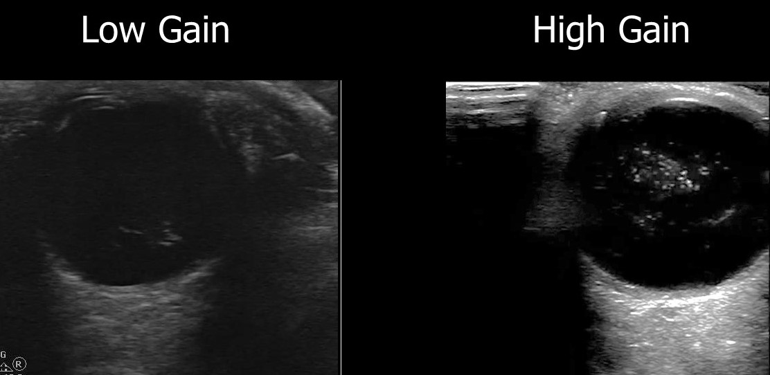
A recent retrospective review looked at how ultrasound image gain levels impacted the accuracy of POCUS for detection of retinal detachment, retinal hemorrhage and posterior vitreous detachment.
They included 383 ED patients who received ocular POCUS and ophthalmology consultations.
Conclusions: The authors found that increasing the gain for ocular POCUS images allowed for increased sensitivity.
Here is an example of a vitreous hemorrhage on ocular POCUS using low gain and high gain.

Chang M, Finney N, Baker J, Rowland J, Gupta S, Sarsour R, Saadat S, Fox JC. Optimal Image Gain Intensity of Point-of-care Ultrasound when Screening for Ocular Abnormalities in the Emergency Department. West J Emerg Med. 2023 May 5;24(3):622-628. doi: 10.5811/westjem.59714.
Category: Ophthamology
Keywords: Optho. (PubMed Search)
Posted: 6/16/2023 by Robert Flint, MD
(Updated: 6/29/2023)
Click here to contact Robert Flint, MD

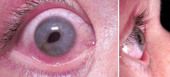
What is this called? What does it indicate? Treatment?
Tear Drop pupil. Globe rupture/corneal laceration. Protect the eye with a commercially available shield (Fox, etc) or if none is available use a paper/sytroform cup that can be cut to length to allow taping in place. Start IV antibiotics and emergency opthamology referal.
1. https://www.jems.com/patient-care/traumatic-eye-injury-management-principl-0/
2. https://eyesoneyecare.com/resources/ophthalmic-emergencies-open-globe-injuries/
Category: Ophthamology
Keywords: corneal perforation (PubMed Search)
Posted: 6/16/2023 by Robert Flint, MD
(Updated: 2/7/2026)
Click here to contact Robert Flint, MD

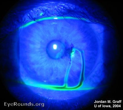
What is this called? What does it indicate?
Seidel Sign. Assoicated with corneal perforation.
https://webeye.ophth.uiowa.edu/eyeforum/atlas/pages/corneal-perforation-seidel-posative-.html
Category: Ophthamology
Keywords: Conjunctivitis (PubMed Search)
Posted: 4/15/2010 by Michael Bond, MD
(Updated: 8/28/2014)
Click here to contact Michael Bond, MD
All to often we see children that are sent to the ED for "Pink Eye" as the school nurse will not allow them back into class unless they are treated with antibiotics. A recent study out of New York identified 4 factors that are associated with low risk (<8% chance) of bacterial (culture postive) conjunctivitis. They are:
An editorial in journal watch comments that if this study can be replicated in other geographic areas we could change the practice of prescribing antibiotics that are not necessary.
Meltzer JA et al. Identifying children at low risk for bacterial conjunctivitis. Arch Pediatr Adolesc Med 2010 Mar; 164:263.
Category: Ophthamology
Keywords: Uveitis, Iritis (PubMed Search)
Posted: 1/16/2010 by Michael Bond, MD
Click here to contact Michael Bond, MD
Iritis is a common diagnosis in the ED, but did you know it was actually a subset of Uveitis.
Uveitis is an inflammation of one or all parts of the uveal tract which consists of the iris, the ciliary body, and the choroid.
The subsets of uveitis are:
Treatment of iritis and uveitis next week.
Emedicine Iritis and Uveitis http://emedicine.medscape.com/article/798323-overview
Category: Ophthamology
Keywords: Sudden Vision Loss (PubMed Search)
Posted: 11/28/2009 by Michael Bond, MD
(Updated: 12/5/2009)
Click here to contact Michael Bond, MD
Some of the causes of acute vision loss are:
Farino GA, Feliciano A, Lorenzo NY. Sudden Visual Loss. eMedicine. Accessed November 27, 2009.
Category: Ophthamology
Keywords: Suden Vision Loss (PubMed Search)
Posted: 11/28/2009 by Michael Bond, MD
(Updated: 2/7/2026)
Click here to contact Michael Bond, MD
Vision loss whether acute or chronic is a common presenting complaint to the ED. This will be the first in a series of pearls on the subject. This pearl will address the nomenclature used by ophthalmology based on the length of vision loss.
• Transient visual obscuration - Episodes lasting seconds. Usually associated with papilledema and increased intracranial pressure.
• Amaurosis fugax - Brief, fleeting attack of monocular partial or total blindness that lasts seconds to minutes
• Transient monocular visual loss or transient monocular blindness - A more persistent vision loss that lasts minutes or longer
• Transient bilateral visual loss - Episodes affecting one or both eyes or both cerebral hemispheres and causing visual loss
• Ocular infarction - Persistent ischemic damage to the eye, resulting in permanent vision loss
Category: Ophthamology
Keywords: MEWDS, White Dot Syndrome (PubMed Search)
Posted: 11/21/2009 by Michael Bond, MD
Click here to contact Michael Bond, MD
MEWDS (Multiple Evanescent White Dot Syndrome)
Multiple Evanescent White Dot Syndrome (MEWDS): 24 y.o. woman with one week duration of central scotoma, OD.
Presented by Sudeep Pramanik, MD, MBA, Hilary A. Beaver, MD http://webeye.ophth.uiowa.edu/eyeforum/cases/37-MultipleEvanescentWhiteDotSyndromeMEWDS.htm
