UMEM Educational Pearls - By Michael Bond

Category: Orthopedics

Title: Spondyl.... Definitions

Keywords: spondyloysis, spondylosis, spondylolistesis, spondylitis (PubMed Search)

Posted: 12/4/2010 by Michael Bond, MD (Updated: 3/29/2024)
Click here to contact Michael Bond, MD

Dr. Corwell covered Spondyloysis in July 2010  https://umem.org/res_pearls_referenced.php?p=1134 but if you are like me you might have trouble remembering the differences between the following terms:

  • Spondyloysis: A unilateral or bilateral defect in the pars interarticularis portion of a vertebrae.  Typically L5 or L4.
  • Spondylosis: is a term referring to degenerative osteoarthritis of the joints between the spinal vertebrae and/or neural foraminae.
  • Spondylolisthesis: describes the anterior displacement of a vertebra or the vertebral column in relation to the vertebrae below. Usually due to spondyloysis or a fracture of the pedicles of the vertebrae.  Can occur anywhere along the vertebral column. Most common at the L4 and L5 level.  For example,  a hangman's fracture is a spondylolisthesis of the C1 vertebra being displaced anteriorly relative to the C2 vertebra.
  • Spondylitis: is an inflammation of the vertebra. As can be seen with ankylosing spondylitis, Pott’s disease or any infection or arthritic disorder of the spine.

Show References


Category: Orthopedics

Title: Evaluation of Potential Intra-Articular Joint Lacerations

Keywords: Methylene Blue, Intra-articular, Joint (PubMed Search)

Posted: 11/21/2010 by Michael Bond, MD
Click here to contact Michael Bond, MD

Evaluation of Potential Intra-Articular Joint Lacerations

Skin and soft tissue injuries in proximity to a joint often prompt concern of whether the injury violated the joint space.  Joint Space involvement is important to exclude as it can lead to septic joints and long term disability.

One easy way to determine if the joint capsule has been violated is to inject methylene blue into the joint and watch to see if any of the methylene blue extravasates through the soft tissue.

Indications for a methylene blue injection include:

  1. Periarticular fracture
  2. Visible joint capsule
  3. Proximity to a joint

There are no absolute contraindications.  Though clearly the procedure does not need to be done when the injury  highly suggests an open joint injury and the patient will require operative debridement and exploration.

To watch a video of a injection head to eMedicine by clicking http://emedicine.medscape.com/article/114453-overview

Show References


Category: Orthopedics

Title: Risk Factors for Spinal Epidural Abscesses

Keywords: Spinal Epidural Abscess (PubMed Search)

Posted: 10/30/2010 by Michael Bond, MD (Updated: 3/29/2024)
Click here to contact Michael Bond, MD

Risk Factors for Spinal Epidural Abscesses

Building on Dr. Corwell's pearl from last week concerning Spinal Epidural Abscess, risk factors for Spinal Epidural Abscesses other than IV drug abuse are:

  1. Diabetes
  2. ESRD
  3. Septicemia
  4. HIV infection
  5. Malignancy
  6. Morbid obesity
  7. Long-term corticosteroid use
  8. Alcoholism
  9. Infection at a distal site
  10. Indwelling catheters
  11. Spinal surgery

The infection can occur via three routes 1) hematogenous spread 2) Direct Extension from a local infection such as osteoomyelitis, and 3) iatrogenic introduction which is thought to be responsible for 14-22% of the cases.  A catheter in the epidural space for more than 2 days has a infection rate of 4.3%.

Show References


Category: Orthopedics

Title: Subungual Hematomas

Keywords: Subungual Hematomas (PubMed Search)

Posted: 10/16/2010 by Michael Bond, MD (Updated: 3/29/2024)
Click here to contact Michael Bond, MD

Subungual Hematomas:

  • Subungual hematomas are collections of blood that form under the nail with injuries to the distal phalanx. 
  • Those that are < 25% of the nailbed can be drained via trephination and heal well.
  • Up to 94%  of subungual hematomas that are are associated with a distal phalanx fracture have a nailed laceration.  It is commonly taught this hematomas should have the nail removed and the nailbed repaired.  However  studies from the 1990's have shown that as long as the nail is attached to the nailbed or paronychia and is not displaced; trephination alone can be done to achieve similar outcomes.

Show References


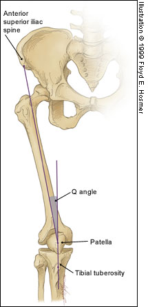
Patellofemoral Syndrome (Chondromalacia Patella)

  • Due to degeneration of the cartilage underneath the patella
  • Patients often present with:
    • A grinding sensation when the knee is extended
    • Pain in the front of the knee that typically worsens after sitting for a long period of time
    • Knee pain that worsens with using stairs, running or when needing to bend the knee deeply (i.e.: squats)
  • Commonly thought to be due to overuse (i.e.: new running program, or marching as in military recruits), but can also be due to anatomic abnormalities like pes planus or a large Q angle.  Ultimate cause is likely to be multifactorial
  • Can be treated with NSAIDs, and limiting activity
  • Physical Therapy that helps to strengthen the quadriceps can help prevent the patella from grinding on the femoral condyles.

Show References


Category: Orthopedics

Title: Pain Control in the Elderly

Keywords: Pain, Geriatrics (PubMed Search)

Posted: 9/18/2010 by Michael Bond, MD (Updated: 3/29/2024)
Click here to contact Michael Bond, MD

Pain Control in the Elderly

  • Narcotic pain relievers are often avoided in the elderly due to the concern of sedation, risk of falls and the concern of them causing delirium.
  • Delirium can cause significant morbidity and mortality and can be difficult to differentiate between the sedation and mild confusion that often occurs with opioid dose escalation.
  • However, delirium has been shown to occur more commonly as a result of the under treatment of pain rather than as an opioid adverse effect.

So the take home lesson for this pearl is that the elderly have a lower risk of delirium if their pain is treated appropriately.

Show References


Adhesive Capsulitis -- Frozen Shoulder

  1. Characterized by pain and loss of motion or stiffness in the shoulder.Normally not seen below the age of 40, affects ~2% of the population and diabetics are at increased risk.
  2. Due to thickening and contracture of the capsule surrounding the shoulder joint.
  3. Can occur after trauma to the shoulder if the shoulder is not moved early enough, but is also know to occur idiopathically.
  4. X-rays are only helpful to rule out other causes of the shoulder pain and are typically normal in Adhesive capsulitis.
  5. Typically will get better on its own over 2-3 years.
    1. Physical Therapy and home exercises aimed at restoring ROM can shorten the duration of pain and stiffness.
    2. Surgery can be done if there is no improvement with medical management and physical therapy.
  6. Prevention strategies include early ROM exercises in those with shoulder injuries especially in the elderly diabetic.

Show References


Category: Orthopedics

Title: Rotator Cuff Tears

Keywords: Rotator Cuff Tears, Chronic, Acute (PubMed Search)

Posted: 8/21/2010 by Michael Bond, MD
Click here to contact Michael Bond, MD

Rotator Cuff Tears:

Four muscles make up the rotator cuff (SITS) which control internal and external rotation of the shoulder and abduct the shoulder.

  1. Supraspinatus
  2. Infraspinatus
  3. Teres Minor
  4. Subscapularis

Tears can be due to acute injuries (falls, heavy lifting, forceful abduction), though the majority (>90%) of rotator cuff tears are chronic in nature and due to subacromial impingement and decreased blood supply to the tendons.

Most patients can be treated with sling immobilization, NSAIDs and referral to sports medicine or orthopaedic surgeons.  Elderly patients should be referred quickly as prolonged immobilization can lead to a frozen shoulder.

Show References


Some common injuries and their board review associated complications

  • Anterior Shoulder Dislocation = Axillary nerve or artery injury
  • Supracondylar Fracture = Brachial Artery injury
  • Posterior Elbow Dislocation = Brachial Artery injury
  • Knee Dislocation = Popiteal Artery Injury and Peroneal and tibial nerve injury
  • Humeral shaft fracture = radial nerve injury
  • Posterior hip dislocation = sciatica nerve injury
  • Anterior hip dislocation = femoral nerve injury

Show References


Diabetic Ketoacidosis Treatment:

  • At least at our academic medical center, we find it very hard to get a DKA patient admitted to an ICU or IMC while they are still in DKA.  Typically, we can correct the acidosis and downgrade them to a floor bed before their ICU/IMC bed is available.
  • Some key points to remember when managing DKA in the ED.
    • The mainstay of treatment for the hyperglycemia initially is IV fluids.
    • Check labs often and replete Magnesium and Potassium early.
    • Insulin should not be started until the potassium is confirmed to be >3.3 mEq/L
    • Patients can still be in DKA even though there glucose is normal.
    • Intravenous insulin must be continued until all the ketones are cleared. 
    • Add D5W or D10 if needed to ensure that their glucose levels stay up but do not stop the insulin.
    • Patients need to receive a long acting insulin (i.e.: Lantus or NPH) 2 hours before the insulin drip is stopped.  Placing a patient only on Sliding Scale Insulin will almost guarantee that they go back into DKA on the floor.
    • Typically you can just restart the patients home long acting insulin, but if you are leary about hypoglycemia if they are not eating well, then give them 3/4 their home dose.

Show References


Category: Toxicology

Title: Anticholinergic or Sympathomimetic

Keywords: anticholinergic, sympathomimetic, pupil (PubMed Search)

Posted: 7/22/2010 by Michael Bond, MD (Updated: 7/24/2010)
Click here to contact Michael Bond, MD

A patient arrives via EMS agitated with VS: P 140, BP 155/100, R 18, T 101F. There is an admitted drug exposure and you examine his eyes which are dilated. You shine the light in the eyes - if the pupil reacts, would that be consistent with anticholinergic or sympathomimetic toxidrome?

Answer: Anticholinergic exposure paralyzes pupillary constrictor muscles and causes dilated pupils that do not react to light. Think about when you go to the eye doctor's office. They put homoatropine in your eyes so that when they look with the slit lamp they can see the retina without interference from pupillary constriction. Sympathomimetic exposure like cocaine activates pupillary dilator muscles, the constrictors are still intact and will give a reflexive constriction to light.  This patient has reactive pupils and by the mere fact is in Baltimore probability dictates a sympathomimetic exposure like cocaine.

Show References


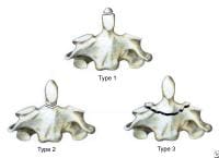
The Salter Harris  Classification System is used in pediatric epiphyseal fractures.  The higher the type of fracture the poorer the prognosis

Some common exam facts about Salter Harris Fractures are:

  1. The type II fracture is the most common.
  2. The small metaphyseal fragment in Salter Harris type II and IV fractures is called the Thurston Holland Sign.
  3. Type III and IV fractures often require open reduction and internal fracture due to the fracture extending into the joint.
  4. Type V fractures may appear normal, but the epiphyseal plate is crushed and the blood supply is interrupted.

The Classification system as listed by Type:

  • Type I: A fracture through the physeal growth plate. Typically can not be seen on x-ray unless they growth plate is widened.
  • Type II: A fracture through the physeal growth plate and metaphysis.
  • Type III: A fracture through the physeal growth plate and epiphysis.
  • Type IV: A fracture through the physis, physeal growth plate and metaphysis.
  • Type V: A crush injury of the physeal growth plate.

A image of the fractures can be found on FP Notebook at http://www.fpnotebook.com/_media/OrthoFractureSalterHarris.jpg

Show References


Category: Gastrointestional

Title: Diverticular Bleeding

Keywords: Diverticular, bleeding, gastrointestinal (PubMed Search)

Posted: 7/3/2010 by Michael Bond, MD
Click here to contact Michael Bond, MD

Diverticular Bleeding

  • Diverticular bleeding is the  most common source of lower GI bleeds and accounts for 17 to 40 percent of cases
  • The most common presentation (80%) is massive painless rectal bleeding. 
  • Patients may have some cramping prior to a bloody bowel movement but otherwise will typically have no abdominal pain.
  • The majority of the cases will resolve spontaneously, but those requiring more than 4 units of Packed Red Blood Cells should be considered for an angiogram or  surgery.
  • Angiography can be used to localize the site of bleeding and embolize the bleeding source. 
  • If embolization fails the patient may require a partial colectomy to treat the bleeding source.

Show References


Category: Orthopedics

Title: Odontoid Fracture

Keywords: Odontoid, fracture (PubMed Search)

Posted: 6/26/2010 by Michael Bond, MD (Updated: 3/29/2024)
Click here to contact Michael Bond, MD

Odontoid Fractures:

There are three types of C2 odontoid fractures:

  1. Type I is an oblique fracture through the upper part of the odontoid process. This fracture is normailly stable and can be treated with a hard cervical collar.
  2. Type II is a fracture occurring at the base of the odontoid as it attaches to the body of C2.  These fractures can be treated surgically, or conservatively with hard collar or a halo brace
  3. Type III fractures occurs when the fracture line extends through the body of the axis. These fractures are normally treated surgically with or without a halo brace.

Odontoid Fractures

Show References


Category: Dermatology

Title: Toxicodendron dermatitis

Keywords: Toxicodendron dermatitis, treatment (PubMed Search)

Posted: 6/19/2010 by Michael Bond, MD (Updated: 3/29/2024)
Click here to contact Michael Bond, MD

Toxicodendron dermatitis:

This is the contact dermatitis caused by the plant genus Toxicodendronm, better known as Poison Ivy.  Here are some types to prevent the dermatitis and how to treat it:

  1. Barrier products like Ivy Block® are on the market that go on like suntan lotion and provides a protective barrier on your skin that prevents the plants urushoil, the toxin responsible for the dermatitis, from making contact with your skin. This can help prevent the dermatitis if you are able to wash the oils off.
  2. Most soaps can not remove urushiol and may actually increase its spread. Several products are on the market, one being Zanfel® , that are a little more effective than water in removing the urushiol which can help to minimize the dermatitis and its spread.
  3. The mainstay of treatment is systemic steroids.  This condition does not do well with a short (5 day) burst therapy and patients will typically get a rebound dermatitis when the burst is complete.  Patients should be placed on a 14 day steroid taper.

Show References


Category: Orthopedics

Title: Calcaneus Fractures

Keywords: Calcaneus Fracture, Bohler Angle (PubMed Search)

Posted: 6/13/2010 by Michael Bond, MD
Click here to contact Michael Bond, MD

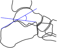
Calcaneus Fractures:

Calcaneus fractures can easily be missed on plain films and the true extent of the injury might not be appreciated until a CT is done.  However, you can increase your change of picking up a calcaneal fracture by evaluating Bohler's Angle. 

Lateral radiographs of the foot are needed to evaluate the Bohler angle.  This is the angle made by drawing a line from anterior process of the calcaneus to the peak of the posterior articular surface and a second one drawn  from the peak of the posterior articular surface to the peak of the posterior tuberosity. (See Picture) The average angle is 25-40°. Angles less than 25' are strongly suggestive of a fracture and the patient should probably get a CT of their foot if there is clinical suspicion.

Bohler's Angle

Show References


Category: Orthopedics

Title: Wound Care

Keywords: Wound Care, Antiseptics (PubMed Search)

Posted: 6/5/2010 by Michael Bond, MD
Click here to contact Michael Bond, MD

Wound Care:

Patients and many providers want to irrigate or wash a wound with an antiseptic solution in order to decrease the risk of infection.  Most studies have shown that irrigation whether with tap water or sterile water is effective enough in reducing bacterial counts in a wound so does adding an antiseptic solution offer any additional benefit.

It turns out that hydrogen peroxide, and iodine based solutions can actually hinder wound healing as they causes delays in the migration and proliferation of fibroblasts at concentrations that are not even bactericidal.  Chlorhexidine, and silver containing antiseptics [i.e.: silver sulfadiazine and silver nitrate] are bactericidal at concentrations that do not affect fibroblasts.

So in the end, if you feel the need to use an antiseptic, use chlorhexidine or a silver containing antiseptic.  The use of hydrogen peroxide and iodine based solutions should be abandoned as they are not even bactericidal at concentrations that have profound affects on the fibroblasts.

Show References


Category: Orthopedics

Title: Septic Arthitis and BioMarkers

Keywords: Septic Arthritis (PubMed Search)

Posted: 5/29/2010 by Michael Bond, MD (Updated: 3/29/2024)
Click here to contact Michael Bond, MD

Septic Arthritis versus Arthritis:

Though CRP and ESR levels are significantly higher in patients that have septic arthritis, a 1998 study showed that there is extensive overlap between patients with septic arthritis  crystal assoicated arthritis that both CRP and ESR have low sensitivity, specificity and predictive values.  Peripherial WBC counts did not differ between the two disease processes..

The morale of the story:  If you are suspecting septic arthritis you need to  perform an arthorcentesis to analysis the synovial fluid.  Systemic biomarkers can not support one diagnosis over the other.

Show References


Category: Orthopedics

Title: Osteomyelitis

Keywords: Osteomyelitis (PubMed Search)

Posted: 5/22/2010 by Michael Bond, MD
Click here to contact Michael Bond, MD

Osteomyelitis:

  • An acute or chronic inflammatory, infectious process of bone.  Can occur via hematogenous spread or direct innoculation of bone.
  • Can be diagnosed on plain radiographs but bony changes might not be evident for 14-21 days.  By 28 days 90% of patients will demonstrate a bony abnormality.
  • Initially plain radiographs will show periosteal elevation. Later cortical or medullary lucencies are seen.
  • Additional tests to help make the diagnosis include:
    • Three phase bone scan: often not practical for the ED.
    • CT Scan: better in areas with complex anatomy [i.e.:spine, pelvis, ,mid and hind foot]
    • MRI: most effective in early detection and to guide surgical approaches.  Sensitivity is estimated at 90-100%.

Show References


Category: Orthopedics

Title: Radial Head Fractures

Keywords: Radial Head, Fracture (PubMed Search)

Posted: 5/16/2010 by Michael Bond, MD (Updated: 3/29/2024)
Click here to contact Michael Bond, MD

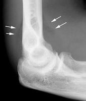
Radial Head Fractures:

Radial head fractures can often be difficult to visualize on plain films especialing Mason Type 1 fractures (see prior pearl on classification system) which are nondisplaced. Often the only sign of a fracture will be a posterior fat pad sign which is always considered to be pathologic.  The posterior fat pad lies outside the synovium of the elbow joint and is normally hidden in the fossa of the distal humerus preventing it from being seen on lateral films of a normal elbow.  Trauma to the elbow that results in a intraarticular fracture (typically a radial head fracture) produces an intra-articular hemorrhage that distends the synovium and displaces the fat out of the fossa, producing the typical triangular radiolucent shadow posterior to the distal end of the humerus.

Show References