65 yo female with breast cancer presents with dyspnea and CXR shown below. Diagnosis? Can anything help clarify the diagnosis?
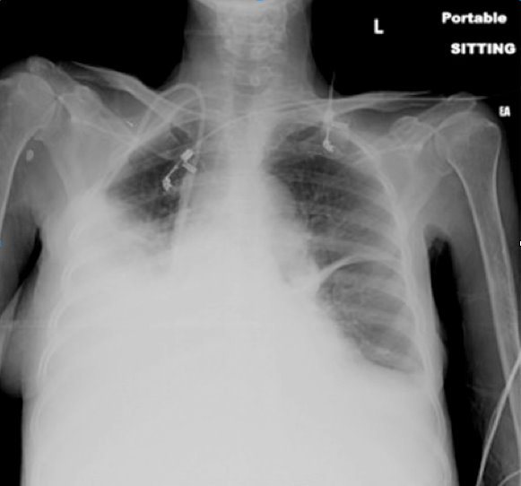
The CXR shows an opaque right hemithorax, consistent with a pleural effusion, a large consolidation, or both. Ultrasound of the lung can clarify this ambiguity.
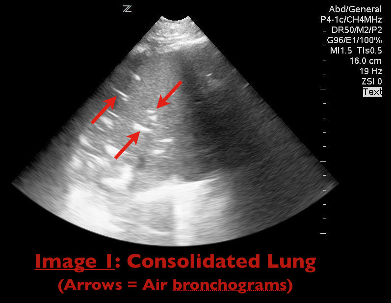
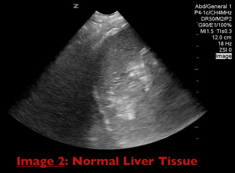
Consolidation of lung parenchyma (e.g., pneumonia) appears isoechoic to liver tissue on ultrasound and air bronchograms appear as hyperechoic areas within the consolidated lung parenchyma (Image 1). Together these findings are termed "hepatization" of the lung as consolidated lung appears similar to liver parenchyma on ultrasound (Image 2).
Durant, A. Nagdev, A. Ultrasound detection of Lung Hepatization. West J Emerg Med. 2010 September; 11(4): 322-323.