Category: Visual Diagnosis
Posted: 6/15/2014 by Haney Mallemat, MD
(Updated: 6/16/2014)
Click here to contact Haney Mallemat, MD
41year-old male without past medical history presents with the image below. What's the diagnosis and what's the most likely causative organism?
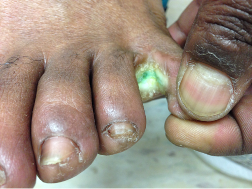
Gram-Negative web-space infection; most common etiologic organism is Pseudomonas
Gram Negative Web-space Infections
Gram-negative infections may occur in toe web-spaces of healthy patients, especially in areas with hyperhidrosis (excessive perspiration) and macerated skin.
Common organisms include Pseudomonas; others include Corynebacterium minutissimum (causes erythrasma, chronic superficial infection of the intertriginous areas of the skin), and other gram-negative bacteria
Woods-light examination may be clinically helpful if Pseudomonas is suspected (fluoresce green) and erythrasma will fluoresce coral red. KOH direct examination for fungal elements may show the presence of dermatophytes or Candida.
Treatment:
Follow me on Twitter (@criticalcarenow) or Google+ (+criticalcarenow)
Wolff, K., & Johnson, R. J. (2009). Fitzpatrick's Color Atlas and Synopsis of Clinical Dermatology. (6th ed., pp. 662-664;698). McGraw-Hill.
http://www.aafp.org/afp/2005/0901/p833.html
http://emedicine.medscape.com/article/1055306-overview
Category: Visual Diagnosis
Posted: 6/8/2014 by Haney Mallemat, MD
(Updated: 6/9/2014)
Click here to contact Haney Mallemat, MD
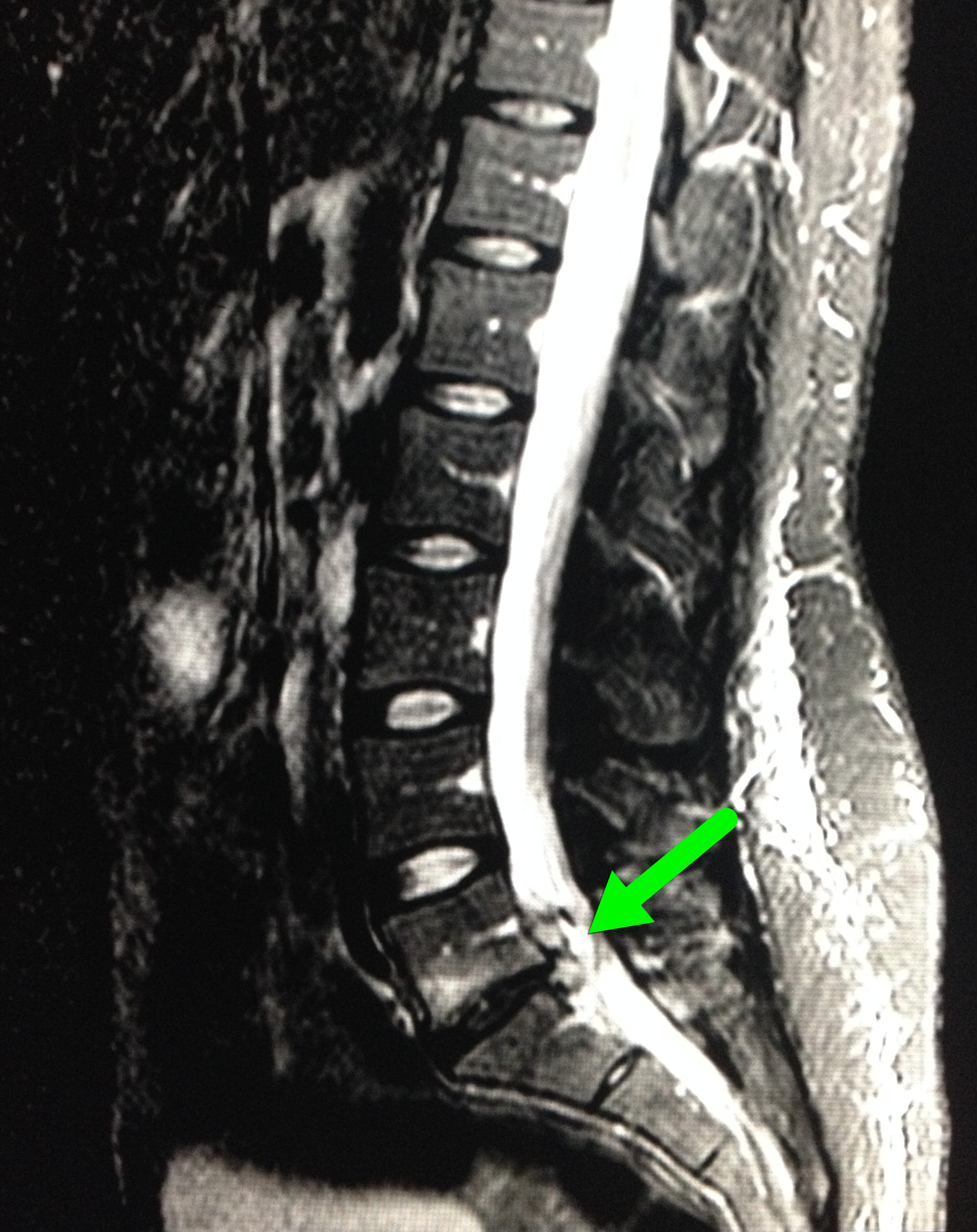
28 year-old female with history of chronic back pain presents with right-foot numbness and inability to move her foot at the ankle joint. What’s the diagnosis and what neurologic finding would you expect to find?
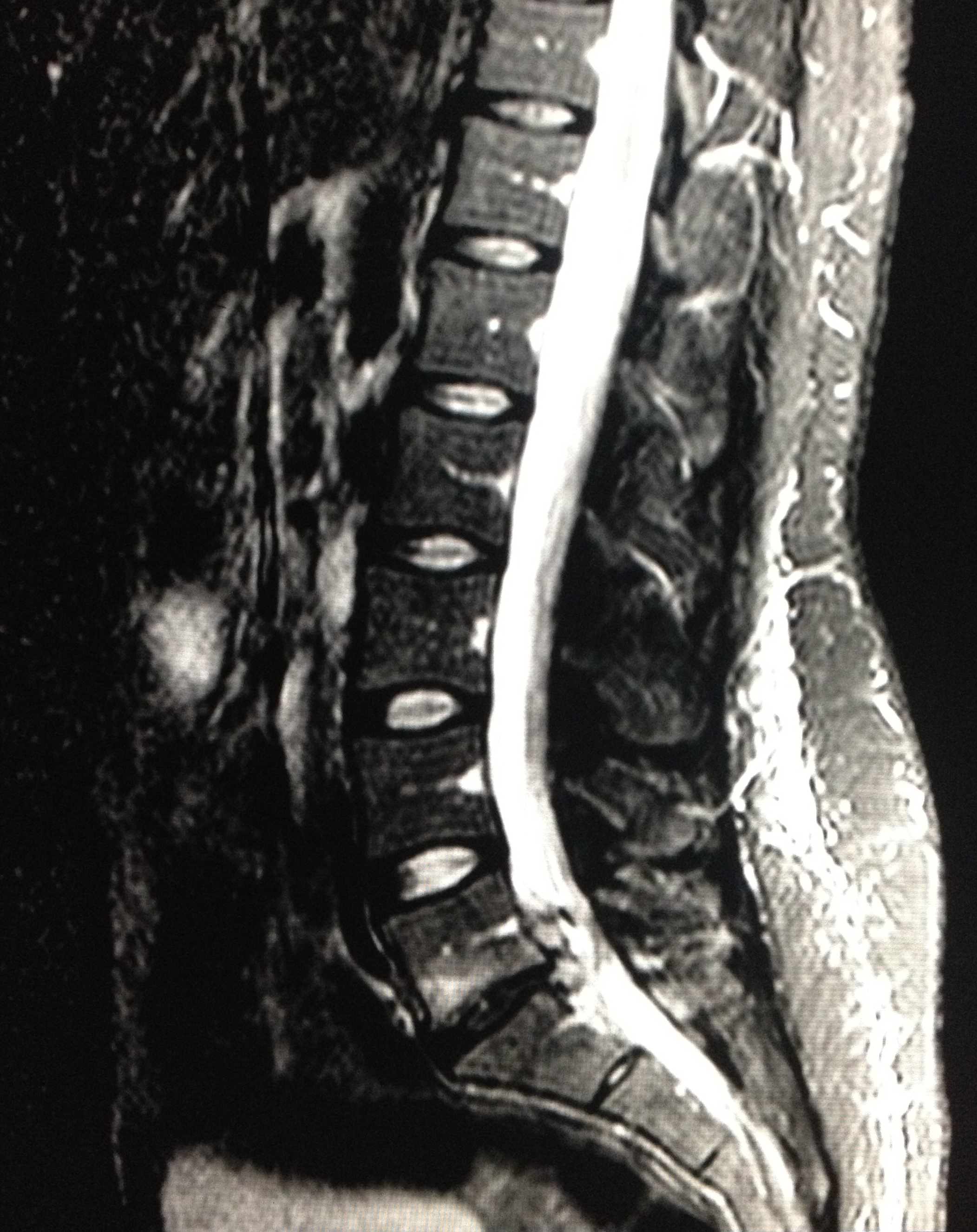
Large disc bulge at L5-S1
Follow me on Twitter (@criticalcarenow) or Google+ (+criticalcarenow)
Category: Visual Diagnosis
Posted: 6/2/2014 by Haney Mallemat, MD
Click here to contact Haney Mallemat, MD
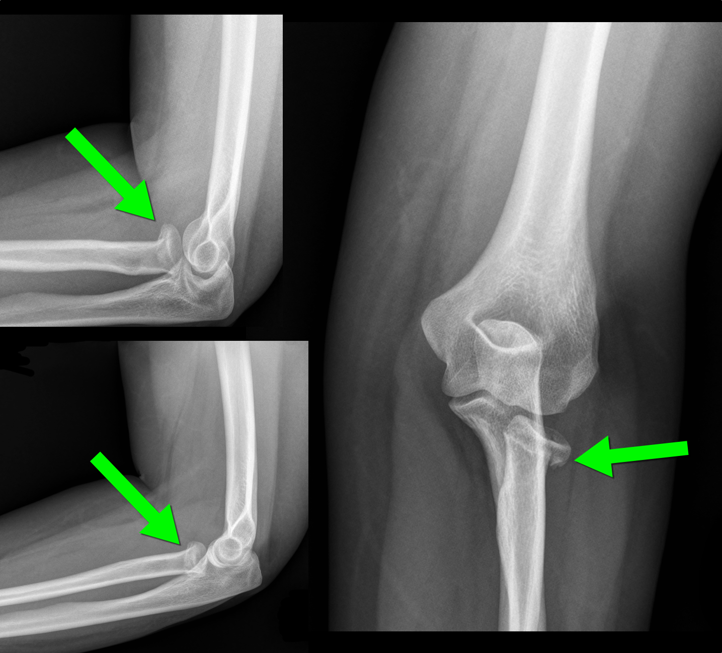
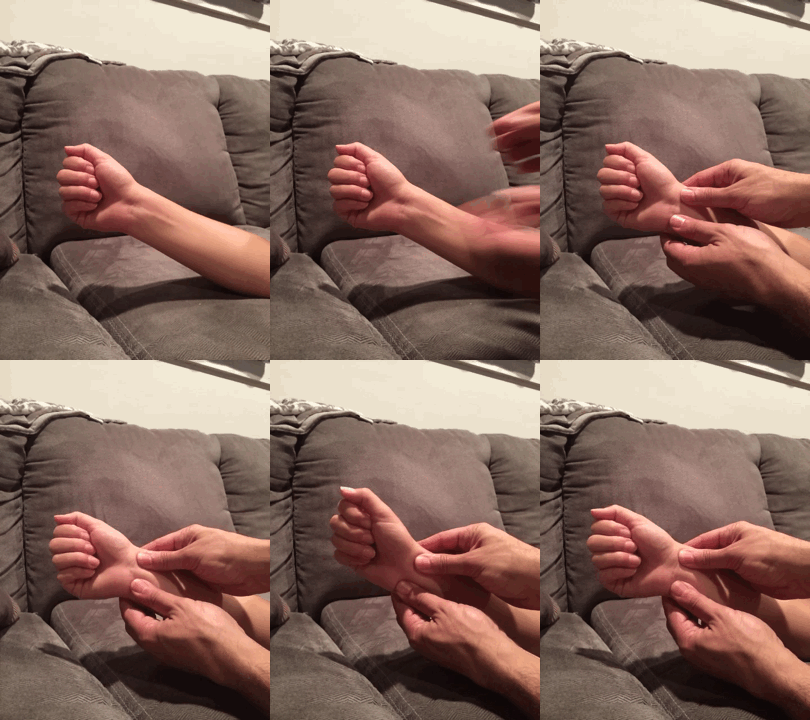
30 year-old female with elbow pain following a fall. What's the diagnosis? (bonus points if you name the fracture with an associated radio-ulnar joint dislocation)
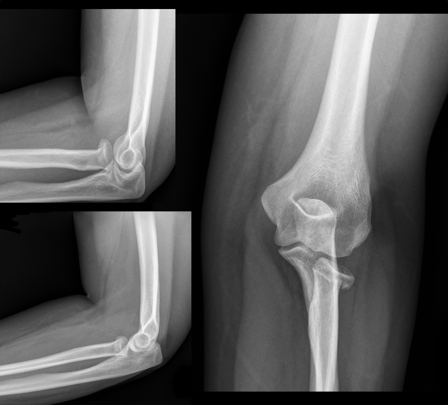
Radial Head Fracture
 d
d
http://www.wheelessonline.com/ortho/radial_head_frx
Follow me on Twitter (@criticalcarenow) or Google+ (+criticalcarenow)
Category: Visual Diagnosis
Posted: 5/26/2014 by Haney Mallemat, MD
Click here to contact Haney Mallemat, MD
Diabetic patient with active intravenous drug use presents with hypotension, fever, and tenderness of right arm. What's the diagnosis and what antibiotic(s) would you start?
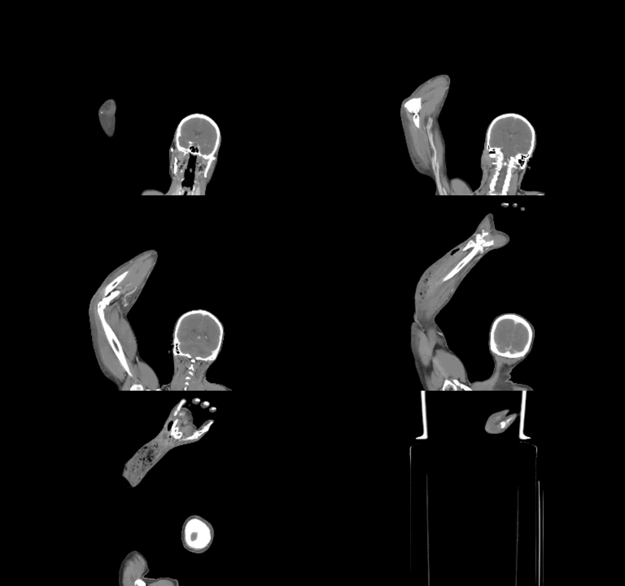
Right arm with necrotizing fasciitis
http://lifeinthefastlane.com/education/ccc/necrotising-fasciitis/
Follow me on Twitter (@criticalcarenow) or Google+ (+criticalcarenow)
Category: Visual Diagnosis
Posted: 5/19/2014 by Haney Mallemat, MD
Click here to contact Haney Mallemat, MD
A patient presents with the rash shown below and is treated with penicillin. Fever, headache, and myalgia develop four hours later. What’s the diagnosis?
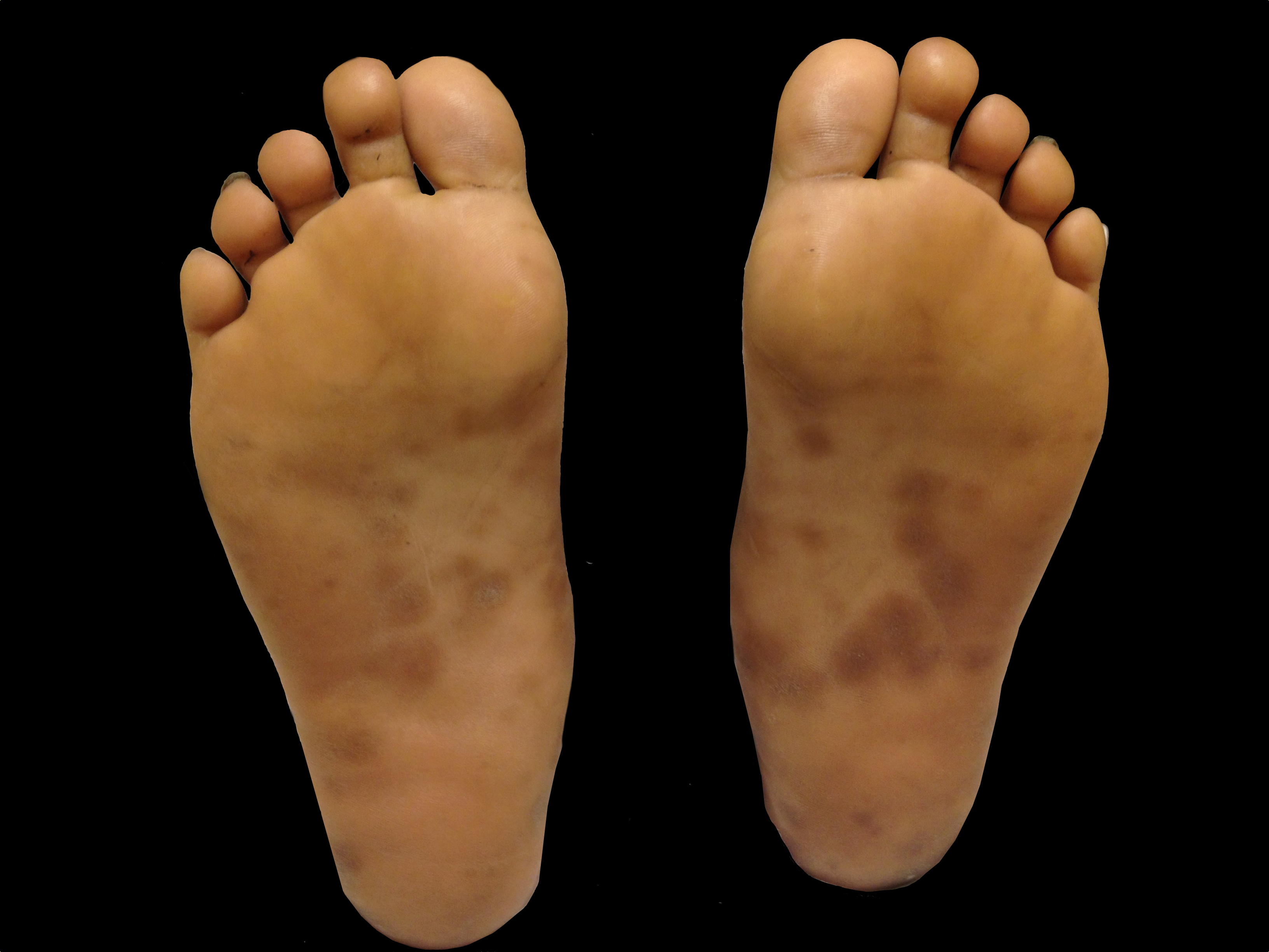
Answer: Jarisch-Herxheimer reaction
Follow me on Twitter (@criticalcarenow) or Google+ (+criticalcarenow)
Category: Visual Diagnosis
Posted: 5/5/2014 by Haney Mallemat, MD
Click here to contact Haney Mallemat, MD
The clip below demonstrates normal right femoral anatomy. The structure with the asterisk is the right common femoral vein and the arrow is pointing to a branch of the right femoral vein. What is the name of the branch and what is its importance during lower extremity ultrasound?
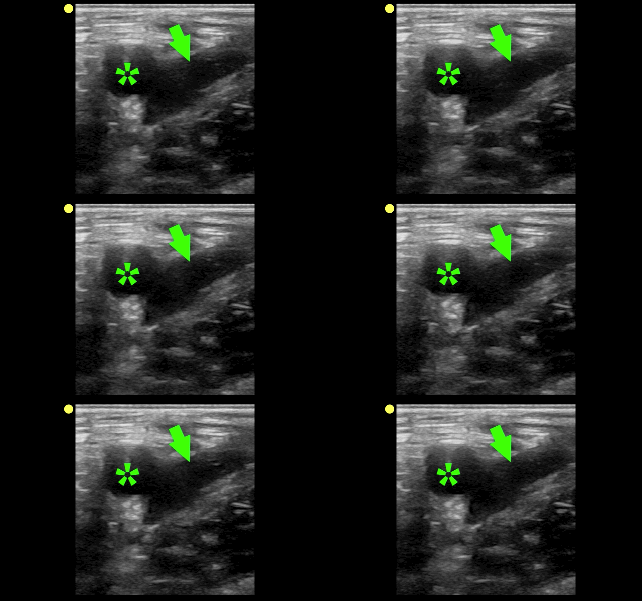
Answer: Greater Saphenous Vein; it is one of the two regions that should be compressed when evaluating for a lower extremity DVT in the Emergency Department (the other is at the trifurcation of the popliteal vein).
Here is a podcast from the Ultrasound Podcast describing the entire bedisde DVT exam http://www.ultrasoundpodcast.com/2011/08/dvt/
Follow me on Twitter (@criticalcarenow) or Google+ (+criticalcarenow)a
Category: Visual Diagnosis
Posted: 4/28/2014 by Haney Mallemat, MD
Click here to contact Haney Mallemat, MD
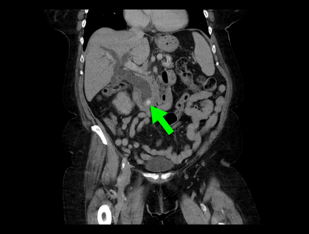
66 year-old female presents with one week of epigastric and right flank pain. Urinalysis was normal. What’s the diagnosis?
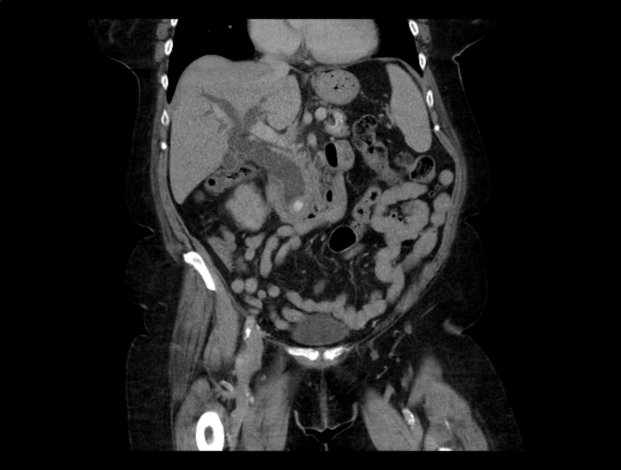
Answer: Choledocholithiasis with pancreatitis secondary to 2.2 cm common bile duct stone
Follow me on Twitter (@criticalcarenow) or Google+ (+criticalcarenow)
Category: Visual Diagnosis
Posted: 4/14/2014 by Haney Mallemat, MD
Click here to contact Haney Mallemat, MD
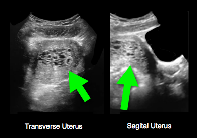
25 year-old female (G1P1) presents with 3 weeks of vaginal bleeding. Her serum beta-HCG is 65,000. Her bedside ultrasound is below; what's the diagnosis?
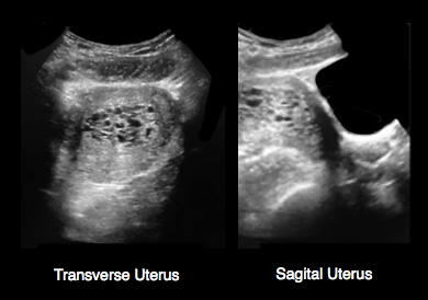
Answer: Hydatidiform mole (molar pregnancy)
Follow me on Twitter (@criticalcarenow) or Google+ (+criticalcarenow)
Category: Visual Diagnosis
Posted: 4/7/2014 by Haney Mallemat, MD
Click here to contact Haney Mallemat, MD
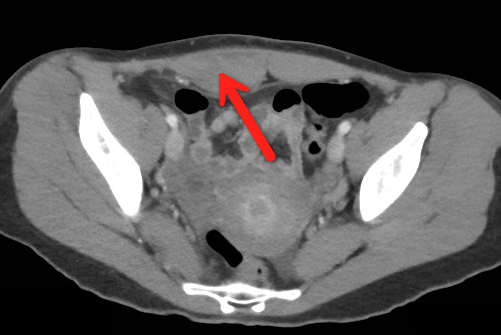
23 year-old female presents complaining of progressive right lower quadrant pain after doing "vigorous" pushups. CT abdomen/pelvis below. What’s the diagnosis? (Hint: it’s not appendicitis)
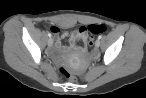
Answer: Rectus sheath hematoma
Rectus Sheath Hematoma (RSH)
Rectus muscle tear causing damage to the superior or inferior epigastric arteries with subsequent bleeding into the rectus sheath; uncommon cause of abdominal pain but mimics almost any abdominal condition.
May occur spontaneously, but suspect with the following risk factors:
Typically a self-limiting condition, but hypovolemic shock may result from significant hematoma expansion.
Follow me on Twitter (@criticalcarenow) or Google+ (+criticalcarenow)
Category: Visual Diagnosis
Posted: 3/31/2014 by Haney Mallemat, MD
(Updated: 4/1/2014)
Click here to contact Haney Mallemat, MD
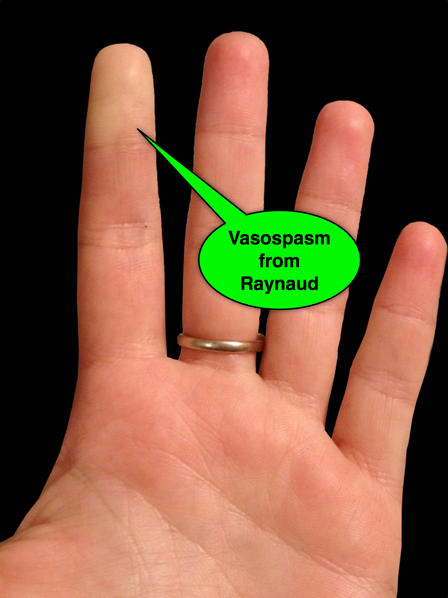
25 year-old female presents with the following. It seems to have occurred spontaneously and spontaneously resolves during her ED evaluation.
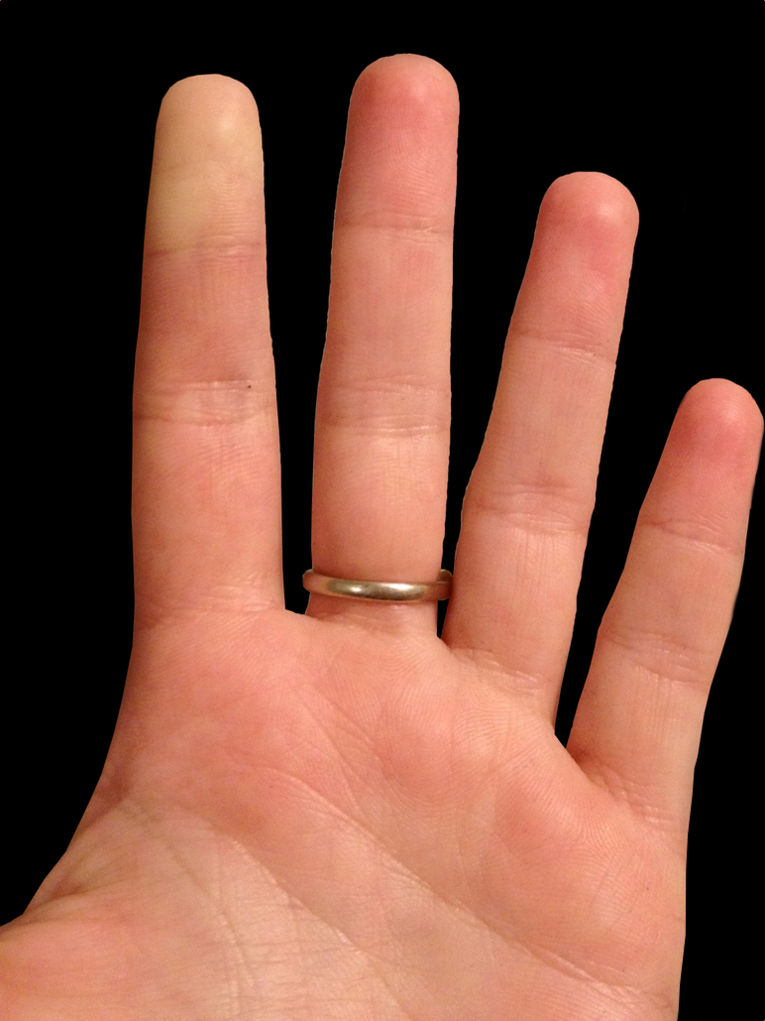
Answer: Raynaud phenomenon
Follow me on Twitter (@criticalcarenow) or Google+ (+criticalcarenow)
Category: Visual Diagnosis
Posted: 3/17/2014 by Haney Mallemat, MD
Click here to contact Haney Mallemat, MD
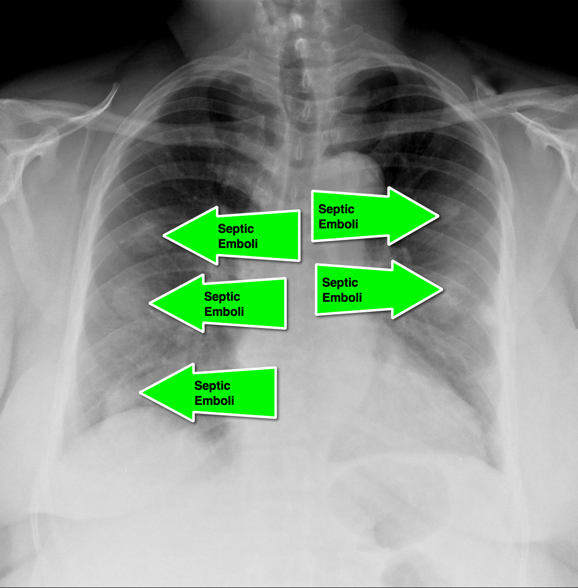
62 year-old male presents with weakness, chills, cough, and malaise. Recently, he had four teeth extracted but felt fine immediately after the extraction. Past medical history includes diabetes and hypertension; CXR is below. What’s the diagnosis?
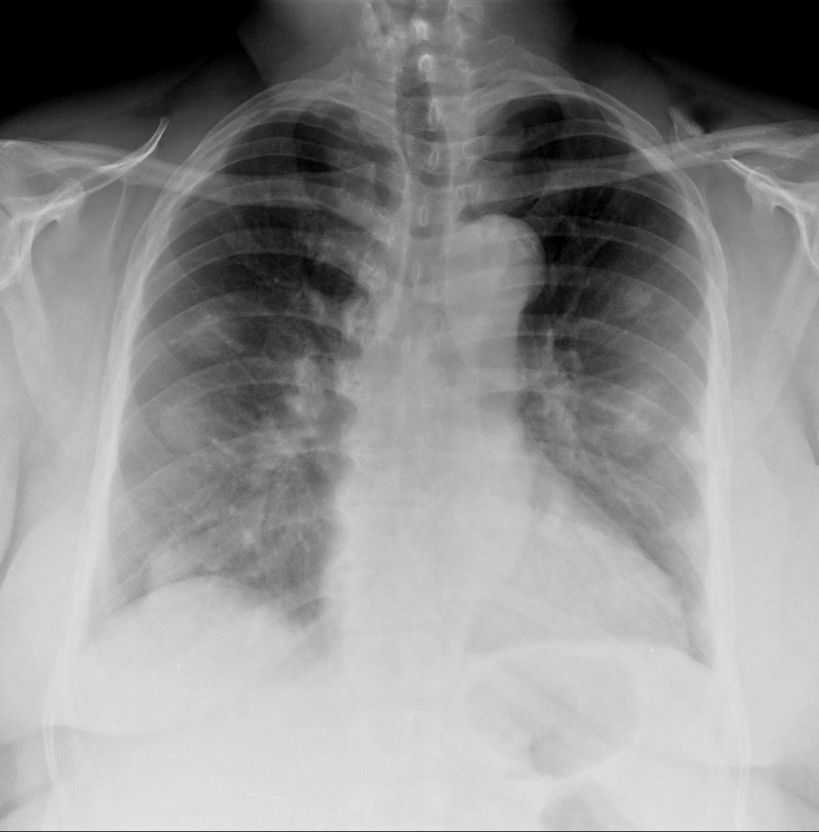
Answer: Septic pulmonary emboli (a.k.a. pulmonary puffballs); he was eventually diagnosed with bacterial endocarditis.
Follow me on Twitter (@criticalcarenow) or Google+ (+criticalcarenow)
Category: Visual Diagnosis
Posted: 3/14/2014 by Haney Mallemat, MD
Click here to contact Haney Mallemat, MD
35 year-old carpet-layer presents with swelling of the superior portion of his knee that has progressively gotten worse over one week. He has no fever and has full range of motion (although pain is worse with movement). The knee is not tender to touch and the area is not erythematous or warm. What's the diagnosis?
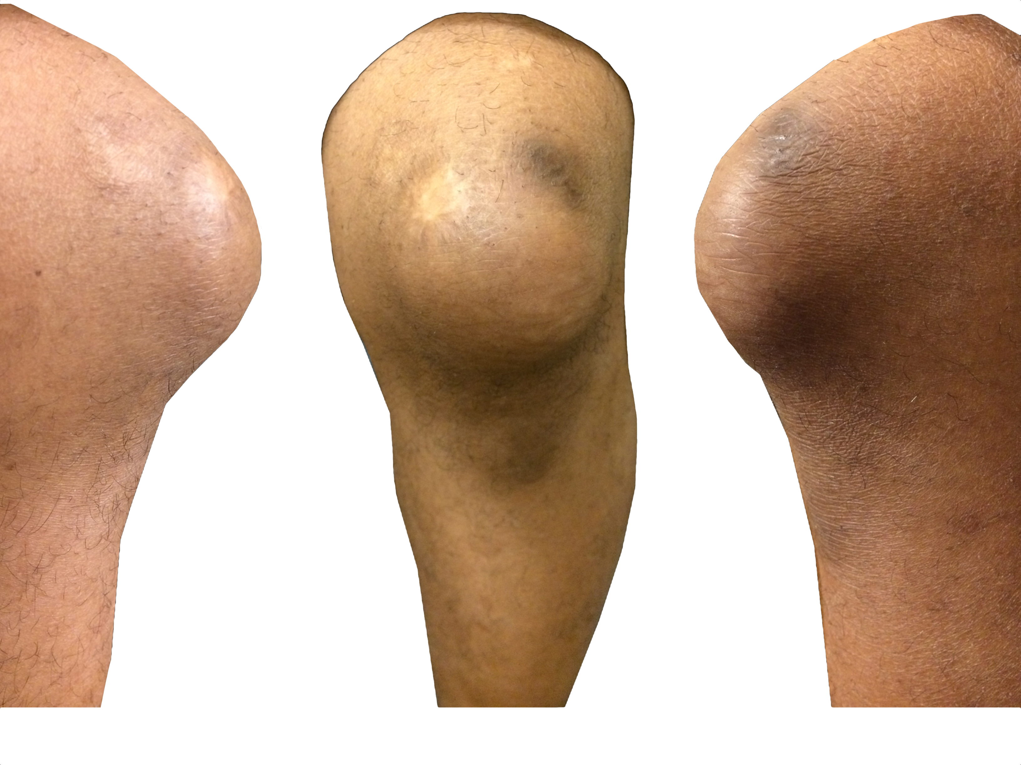
Answer: Pre-patellar bursitis
Follow me on Twitter (@criticalcarenow) or Google+ (+criticalcarenow)
Category: Visual Diagnosis
Keywords: international, global, hypoxia, clubbing (PubMed Search)
Posted: 3/12/2014 by Andrea Tenner, MD
Click here to contact Andrea Tenner, MD
What is this physical finding?
Answer: Clubbing
Differential Diagnosis of Clubbing in Children:
Clubbing was first documented by Hippocrates in a patient with empyema. It has been associated with cardiopulmonary disease, but also found in other disease processes such as hepatobiliary, infectious, and endocrine diseases. It can also occur without an underlying pathology. The pathophysiology behind these physical findings remain unknown. The workup and treatment should be aimed at managing the underlying cause.
University of Maryland Section for Global Emergency Health
Author: Van Pham, MD
Schwartz, Robert. Clubbing of the Nails. 3.12.14. http://emedicine.
Category: Visual Diagnosis
Posted: 3/3/2014 by Haney Mallemat, MD
Click here to contact Haney Mallemat, MD
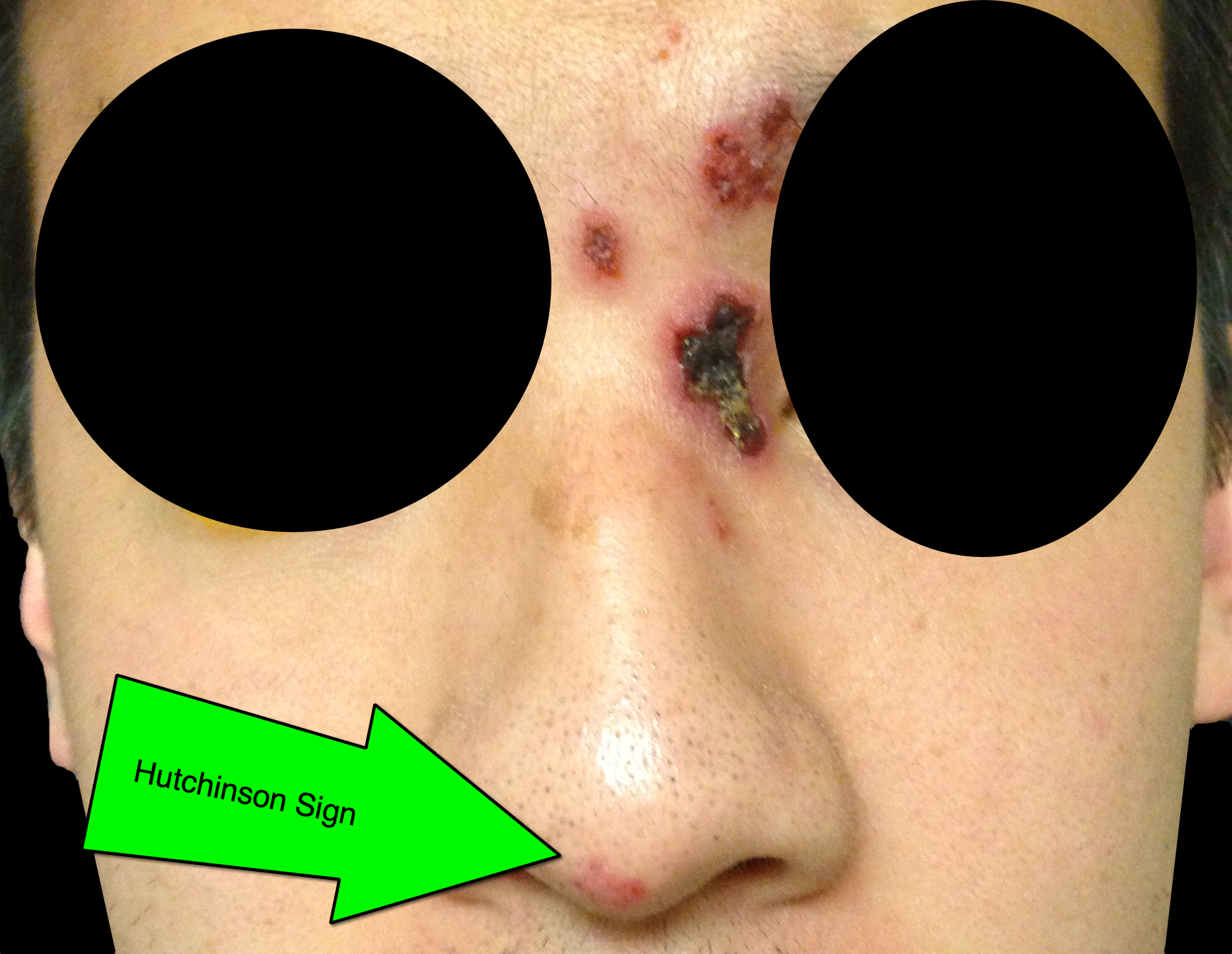
32 year-old male presents with the following. What's the diagnosis?
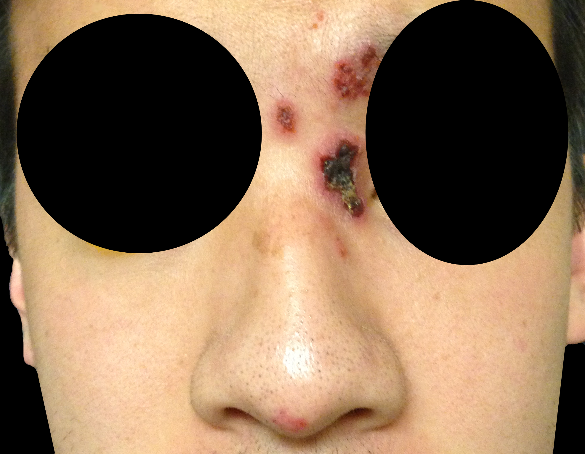
Answer: Hutchinson's Sign
Herpes zoster ophthalmicus (HZO)
Follow me on Twitter (@criticalcarenow) or Google+ (+criticalcarenow)
Category: Visual Diagnosis
Posted: 2/24/2014 by Haney Mallemat, MD
Click here to contact Haney Mallemat, MD
50 year-old with facial weakness and dysarthria. What's the diagnosis?
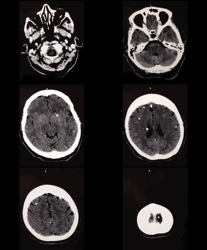
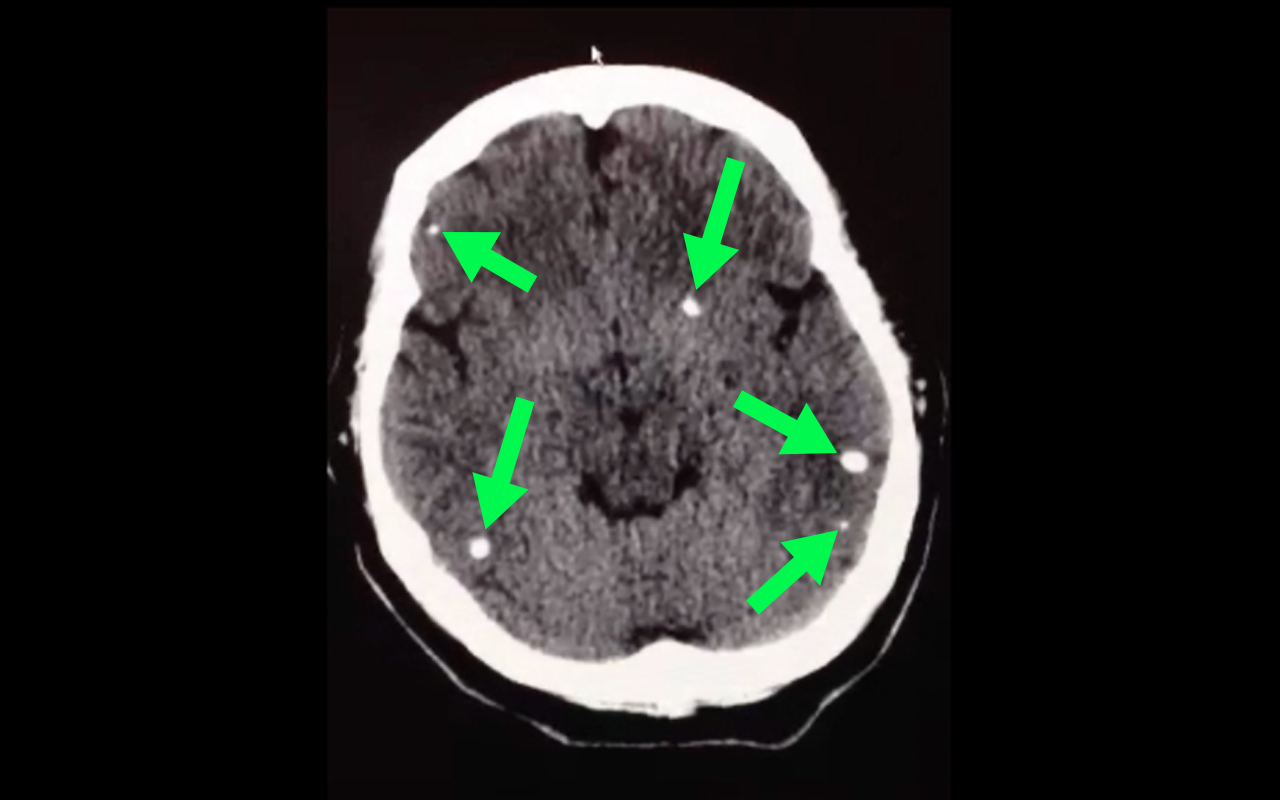
Stroke secondary to neurocysticercosis
Neurocysticercosis
Neurocysticercosis occurs secondary to ingestion of eggs from the Taenia solium tapeworm; ingestion typically occurs from ingestion of contaminated food.
Most patients are asymptomatic but symptoms of neurocysticercosis may include
Diagnosis is made via imaging such as CT scan; findings may vary depending on what stage of life the parasite is in (e.g., larvae); lumbar puncture should also be performed if the diagnosis is suspected.
Management should be symptomatic (e.g., seizures receive anticonvulsants) as well as eradication of the active parasites using steroids followed by albendazole (anti-parasitic drug)
Follow me on Twitter (@criticalcarenow) or Google+ (+criticalcarenow)
Category: Visual Diagnosis
Posted: 2/17/2014 by Haney Mallemat, MD
Click here to contact Haney Mallemat, MD
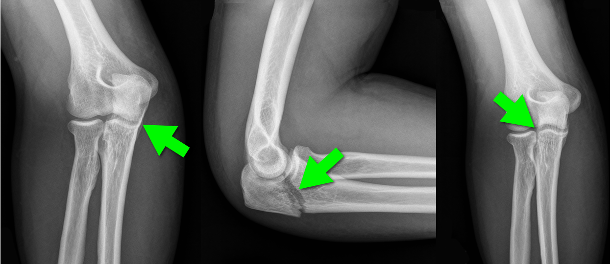
44 year-old construction worker fell off a ladder and presents with elbow pain. What's the diagnosis and what is the most commonly associated nerve injury?
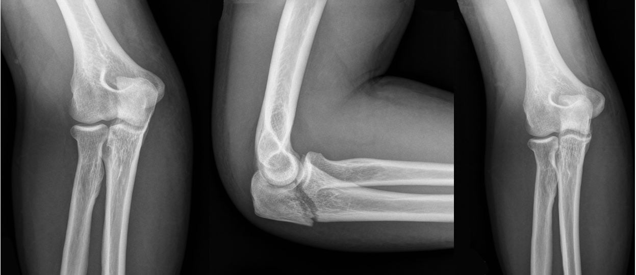
Olecranon fracture; the ulnar nerve is most commonly injured
Pearls for Olecranon Fracture
Follow me on Twitter (@criticalcarenow) or Google+ (+criticalcarenow)
Category: Visual Diagnosis
Posted: 2/9/2014 by Haney Mallemat, MD
(Updated: 2/10/2014)
Click here to contact Haney Mallemat, MD
25 year-old male presents after falling off his bicycle. He complains of pain in his right-hand (he is right-hand dominant). What's the diagnosis?
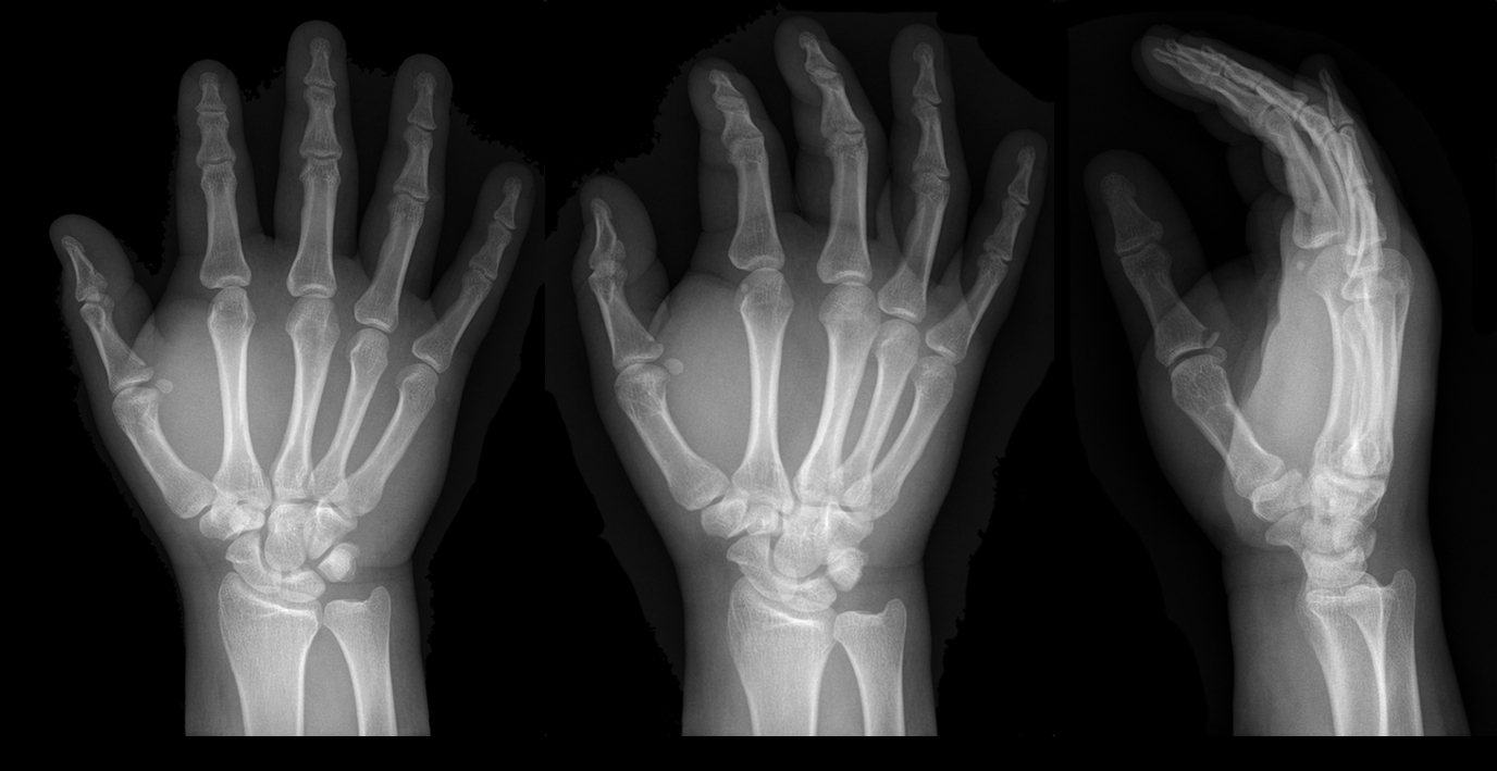
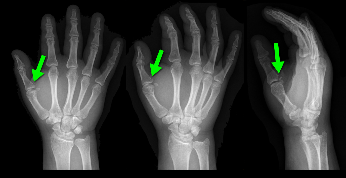
Answer: Gamekeeper's thumb or Skier's thumb
Injury to the ulnar collateral ligament (UCL) secondary
Called “Gamekeeper Thumb” because injury was originally described in gamekeepers who job was to break the necks of captured game (or animals). The act of breaking necks led to a repetitive stress and injury secondary to the valgus forces on the thumb.
UCL can become torn or avulsed from the insertion on the proximal phalanx; the UCL is an important stabilizer of the thumb; improper management can lead to future disability.
Treatment is with a short-arm thumb-spica cast and follow up with a hand surgeon to determine whether management will be conservative or operative.
Chuter GS, Muwanga CL, Irwin LR (June 2009). "Ulnar collateral ligament injuries of the thumb: 10 years of surgical experience". Injury 40 (6): 652–6 Wheeless' Textbook of Orthopaedics Online: Surgical treatment of acute gamekeeper's thumb
Follow me on Twitter (@criticalcarenow) or Google+ (+criticalcarenow)
Category: Visual Diagnosis
Posted: 2/3/2014 by Haney Mallemat, MD
Click here to contact Haney Mallemat, MD
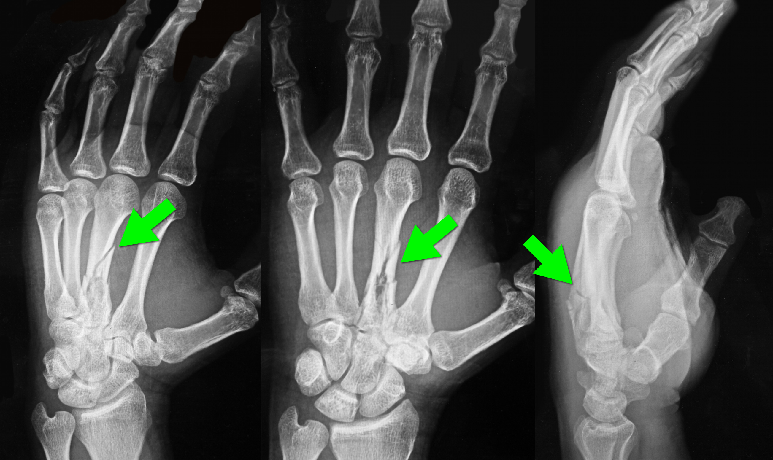
34 year-old left-hand dominant male sustained injury to left hand after his pressurized greasing-gun discharged into the palm of his hand. He has a small lac to the hand but is in extreme pain. On exam his hand is very puffy and he is neurovascularly intact (XR below) What is the next step in management?
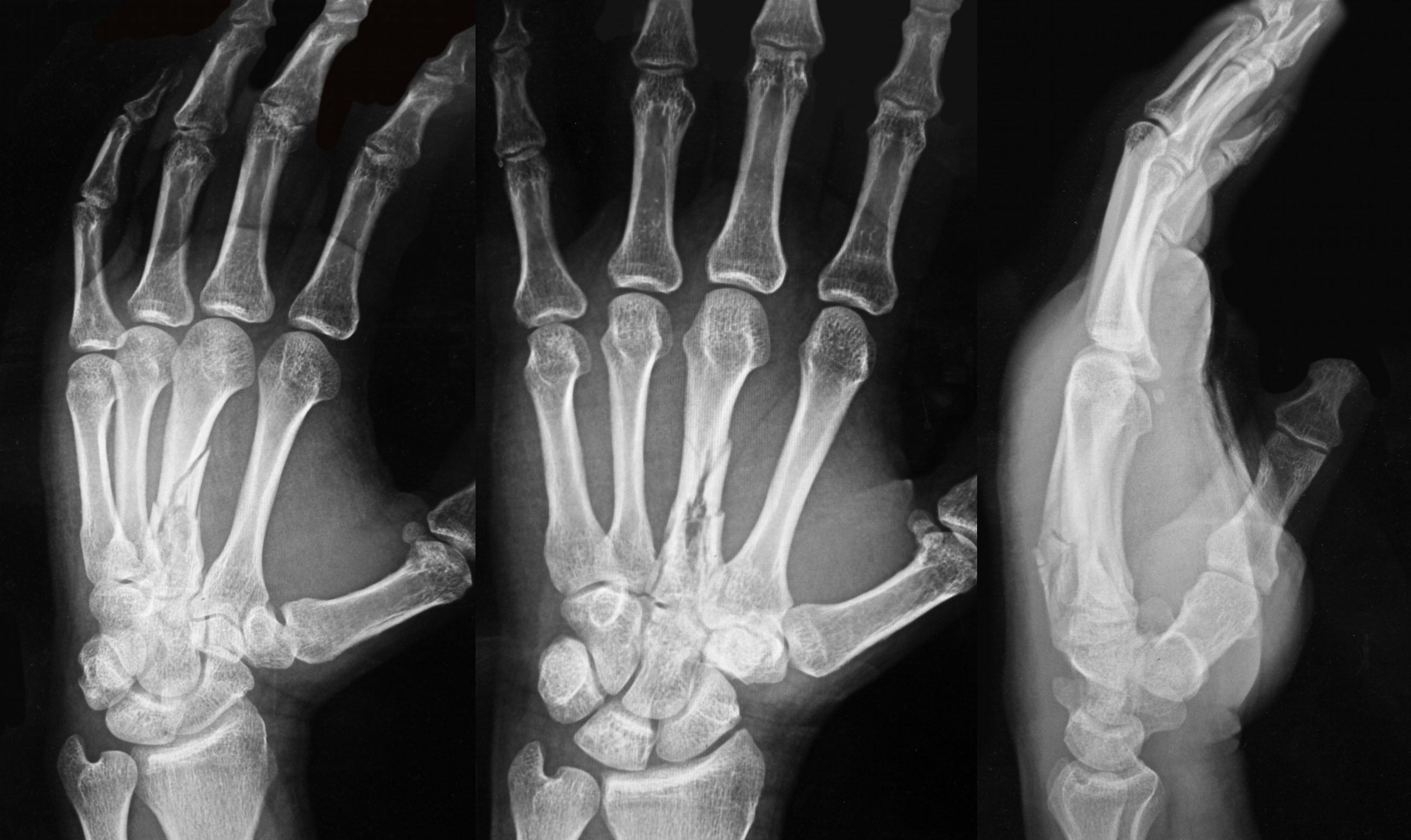
Metacarpal fracture; emergent surgical consult is required
High-Pressure Injection Injuries (HPI) injuries
HPI devices (e.g., grease, paint guns, etc.) may cause injury when the device accidently discharges, typically into the operator’s dominant hand during attempts to clean the nozzle.
Injuries may initially appear benign, however injuries should be considered surgical emergencies because they often require debridement / washout of materials injected into deeper tissues.
Tissue damage is multi-factorial and includes direct injury (e.g., high-pressure tissue damage, vascular compression, etc.) and indirect injury from material injected into tissues (e.g., chemical inflammation / injury, granuloma formation, deep-tissue infections, etc.).
Radiographs may assist surgical planning by identifying subcutaneous air, debris, or unanticipated fractures.
Treatment:
Follow me on Twitter (@criticalcarenow) or Google+ (+criticalcarenow)
Category: Visual Diagnosis
Posted: 1/27/2014 by Haney Mallemat, MD
Click here to contact Haney Mallemat, MD
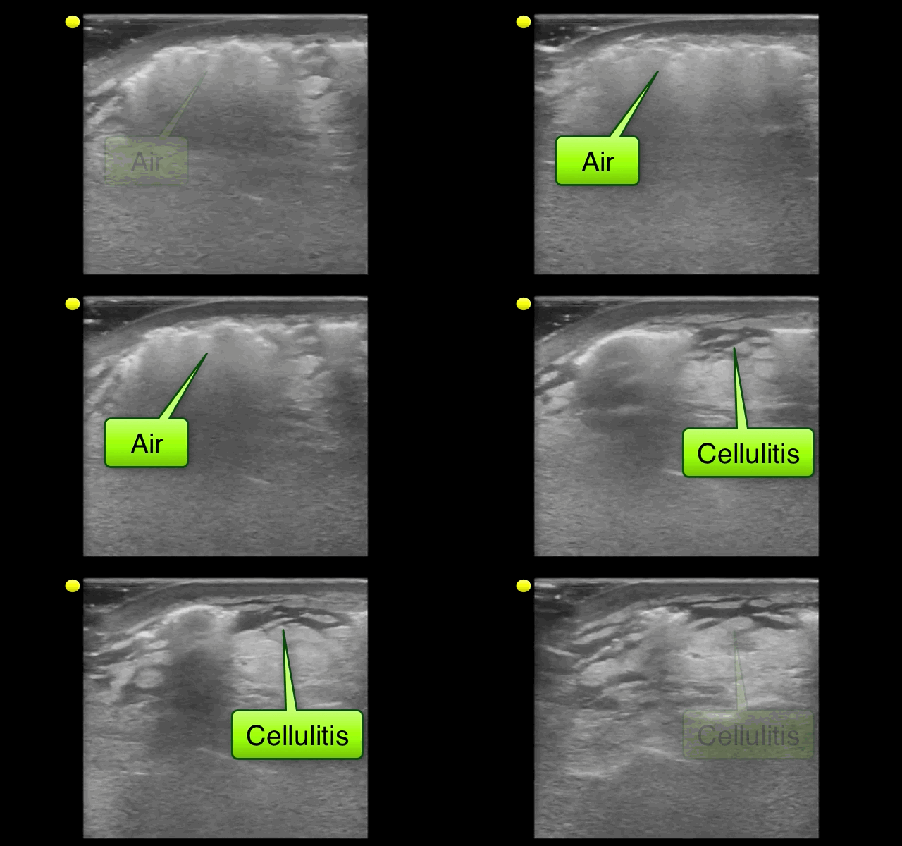
32 year-old with diabetes presents with fever, erythema, and warmth of his lower extremity; his leg is not particularly painful. He is diagnosed with cellulitis, started on antibiotics, and admitted to the hospital. While boarding in the Emergency Department he becomes rigorous and hypotensive. An ultrasound of his cellulitis is performed and is shown below. What’s the diagnosis?
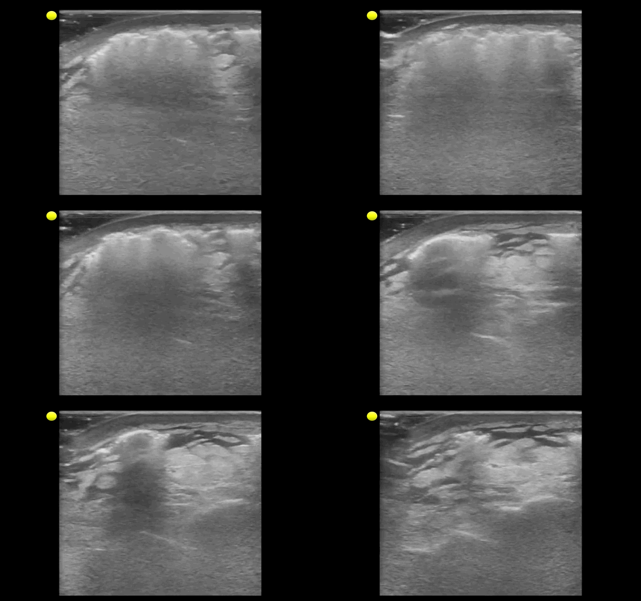
"Cobblestoning" of subcutaneous tissue consistent with cellulitis. There are also areas of subcutaneous air raising the concern for necrotizing fasciitis.
Special thanks to Dr. Mike Mallin from the Ultrasound Podcast for the clip. Want to see more amazing ultrasound clips? Check out Sonocloud
Follow me on Twitter (@criticalcarenow) or Google+ (+criticalcarenow)
Category: Visual Diagnosis
Posted: 1/20/2014 by Haney Mallemat, MD
Click here to contact Haney Mallemat, MD
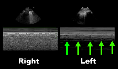
50 year-old male intubated for respiratory distress. Ultrasound is used post-intubation to confirm tube placement and the following images are obtained. What's the diagnosis?
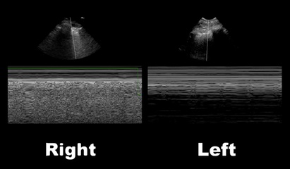
Right main-stem intubation as demonstrated by presence of lung-pulse on the left side
Lung-Pulse
Follow me on Twitter (@criticalcarenow) or Google+ (+criticalcarenow)
