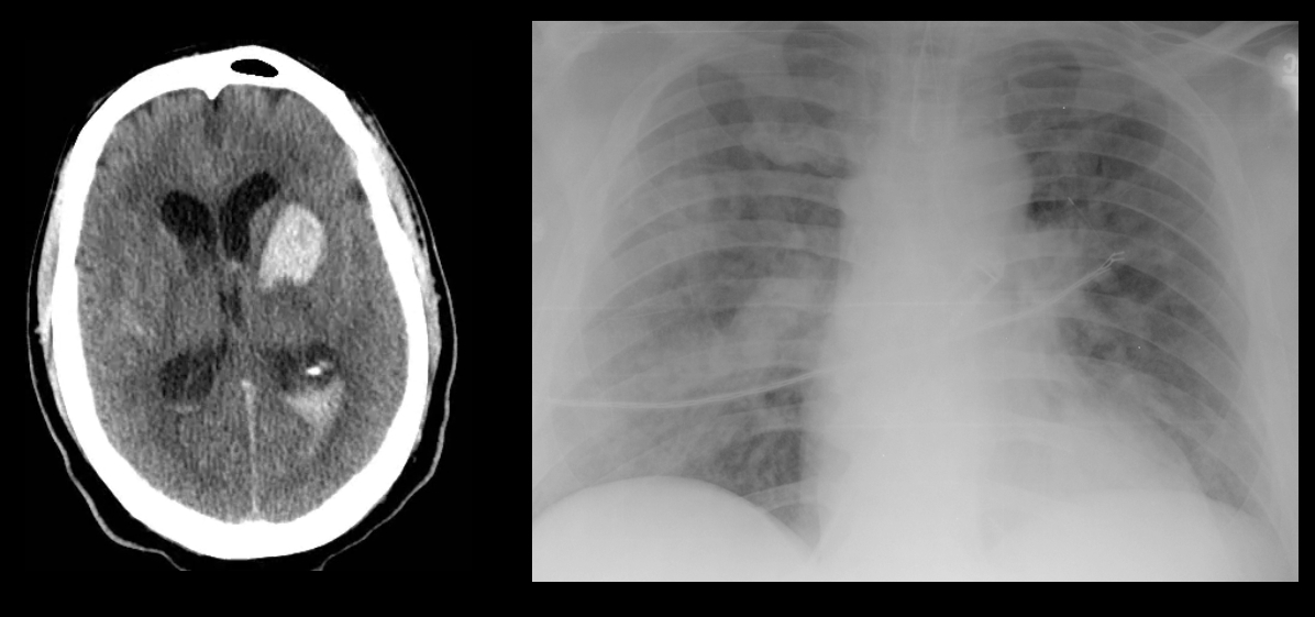
UMEM Educational Pearls - Critical Care

Category: Critical Care

Title: Cerebral Salt Wasting Syndrome vs. Syndrome of Inappropriate ADH Secretion.

Keywords: SIADH, CSW, syndrome of inappropriate adh, cerebral salt wasting, hyponatremia, neurosurgery (PubMed Search)

Posted: 8/30/2010 by Haney Mallemat, MD (Emailed: 8/31/2010) (Updated: 4/27/2024)
Click here to contact Haney Mallemat, MD

Hyponatremia plagues many neurosurgical patients due to the syndrome of inappropriate secretion of ADH (SIADH) or the cerebral salt wasting syndrome (CSW). Both diseases may appear similar (hyponatremia, increased urine osmolarity, increased urine sodium, normal adrenal, renal and thyroid function), but there is one BIG difference. Patients with SIADH are euvolemic or hypervolemic (excess ADH causes fluid retention) whereas patients with CSW are fluid depleted (impaired renal handling of sodium and water). To differentiate, look for signs of hypovolemia: orthostatics, dry mucus membranes, hemoconcentration, pre-renal azotemia, and/or hemodynamics (IVC collapse anyone?).

Bottom line: Distinguish SIADH from CSW because the treatments are exact opposites:

SIADH: Fluid restrict

CSW: Give water and salt (i.e., 0.9% saline)

Show References


Category: Critical Care

Title: Hemostatic Therapy for ICH

Posted: 8/24/2010 by Mike Winters, MD (Updated: 4/27/2024)
Click here to contact Mike Winters, MD

Hemostatic Therapy for ICH - Updated Guidelines

  • The AHA/ASA just published updated guidelines for the diagnosis and treatment of acute spontaneous intracerebral hemorrhage (ICH).
  • Regarding hemostatic therapy, new/revised recommendations from the 2007 AHA/ASA guidelines include:
    • Patients with severe thrombocytopenia or factor deficiency should receive platelets or factor replacement
    • Patients with ICH due to oral anticoagulants (warfarin) should receive intravenous vitamin-K and vitamin-K dependent factor replacement
      • Prothrombin complex concentrates (PCCs) are being increasingly used and are considered a reasonable alternative to FFP.  To date, studies have not shown improved outcome with PCCs.
      • Recombinant factor VIIa (rFVIIa) is not recommended as a sole agent for warfarin-related ICH
    • rFVIIa is not recommended in unselected patients
    • Usefulness of platelet transfusions for patients using antiplatelet medications is unclear and currently investigational.

Show References


Category: Critical Care

Title: Drug-Induced Hypophosphatemia

Posted: 8/10/2010 by Mike Winters, MD (Updated: 4/27/2024)
Click here to contact Mike Winters, MD

Drug-Induced Hypophosphatemia

  • Hypophosphatemia is seen in almost 30% of critically ill patients.
  • As discussed in a prior pearl, hypophosphatemia can result in respiratory failure along with cardiac and neurologic abnormalities.
  • Although common ED causes of hypophosphatemia include sepsis, hypothermia, and dialysis, don't forget about medications.
  • Medications that can cause significant hypophosphatemia in the critically ill (along with their mechanism) include:
    • Decreased GI intake: antacids, sucralfate
    • Transcellular shift: aspirin overdose, albuterol, catecholamines, insulin, and bicarbonate
    • Increased urinary excretion: diuretics, acetaminophen overdose, and theophylline overdose

Show References


Hypocapnia and Brain Injury

  • Hypocapnia indirectly reduces cerebral blood volume through reductions in arterial cerebral blood flow.
  • Despite its continued and frequent use, hypocapnia can actually aggravate cerebral hypoxia through reductions in oxygen supply and increases in cerebral oxygen demand.
  • In addition to inducing further cerebral injury, hypocapnia can cause deleterious effects on the heart, lung, and GI tract.
  • To date, there is no evidence that hypocapnia improves outcome in patients with traumatic brain injury or acute stroke.
  • Induced hypocapnia in critically ill ED patients with acute brain injury should primarily be reserved for those with imminent brain herniation.

Show References


Drug-Induced Thrombocytopenia

  • Thrombocytopenia is common in critically ill patients and is associated with increased mortality.
  • Up to 25% of critically ill patients will develop thrombocytopenia as a result of a medication, termed drug-induced thrombocytopenia (DIT)
  • Antibiotcs are a common, yet infrequently recognized, cause of DIT.
  • Antibiotics reported to cause DIT include linezolid, vancomycin, trimethoprim/sulfamethoxazole, and the beta-lactams.
  • In fact, piperacillin/tazobactam has been associated with DIT more frequently than any other penicillin. 

Show References


Category: Critical Care

Title: ICU Acquired Weakness

Posted: 7/19/2010 by Mike Winters, MD (Emailed: 7/20/2010) (Updated: 4/27/2024)
Click here to contact Mike Winters, MD

ICU Acquired Weakness

  • ICU acquired weakness (ICU-aw) is a general term that refers to the weakness that develops in critically ill patients during the course of their illness - especially in patients with sepsis and those receiving mechanical ventilation.
  • ICU-aw is an very common complication of critical illness that can develop within hours and has been shown to increase the duration of mechanical ventilation and ICU/hospital LOS.  Observational studies have also reported an association with mortality.
  • Risk factors associated with ICU-aw include medications (neuromuscular blocking agents, corticosteroids), hyperglycemia and immobility.
  • For the critically ill ED patient, current recommendations suggest limiting the administration of neuromuscular blocking agents and corticosteroids, when possible.

Show References


Drug-Drug Interactions in the Critically Ill

  • Critically Ill ED patients are at risk for drug-drug interactions (DDIs) due to altered organ function, polypharmacy, and altered drug kinetics.
  • DDIs involving the cytochrome isoenzyme CYP3A4 are of particular importance.
  • CYP3A4 inhibitors, such as macrolides and azoles (fluconazole, voriconazole), can cause serious DDIs when given concomitantly with meds that are a subtrate for CYP3A4 - midazolam, cyclosporine, tacrolimus, diltiazem, amiodarone.
  • Pay particular attention to your transplant patients, as administration of an azole can result in significant cyclosporine or tacrolimus toxicity.

Show References


Asthma, Peak Pressures, and the Ventilator

  • In previous pearls, we have highlighted ventilator settings for the asthmatic, along with the differences between peak and plateau pressure measurements.
  • When ventilating the asthmatic, pay attention to the ventilator settings placed by your respiratory therapist.
  • In general, the respiratory therapist will set the ventilator to stop delivering tidal volumes when the peak pressure exceeds 40-60 cm H2O.
  • For asthmatics, this practice can result in very low tidal volumes.
  • Thus, peak pressure limits must be set higher.
  • As you know, high peak pressures have not been shown to be injurious, provided that the plateau pressure remains < 30 cm H2O

Show References


Category: Critical Care

Title: Acidosis and Intubation

Posted: 6/29/2010 by Mike Winters, MD (Updated: 4/27/2024)
Click here to contact Mike Winters, MD

Pre-existing acidosis and mechanical ventilation

  • Not surprisingly, many critically ill ED patients often develop a metabolic acidosis.
  • To compensate, patients hyperventilate, thereby producing a respiratory alkalosis.
  • When these patients require intubation and mechanical ventilation, be sure to provide the same level of respiratory compensation when setting the ventilator. 
  • Failing to provide a rate sufficient to compensate for the pre-intubation acidosis leads to a rapid drop in pH, bradycardia and eventually asystole.
  • In general, rates can be increased to about 30-35 breaths per minute, after which auto-PEEP becomes problematic.

Show References


Acute renal failure occurs in 1-25% of critically ill patients, with an associated mortality of 28 - 90%. 

The RIFLE Criteria represent the first consensus definition of acute renal failure used to classify critically ill patients as to their kidney function.  Notably, we use the worst possible classification according to the criteria, which measures either serum creatinine, urine output or both. 

Show References


Attachments

1006221408_RIFLE.jpg.doc (103 Kb)


Category: Critical Care

Title: Hypotension and MV

Posted: 6/15/2010 by Mike Winters, MD (Updated: 4/27/2024)
Click here to contact Mike Winters, MD

Hypotension after intubation and initiation of mechanical ventilation

  • Approximately 25-30% of patients develop hypotension after intubation and initiation of mechanical ventilation (MV).
  • Although the literature is not robust, risk factors for hypotension after initiation of MV include:
    • hypotension prior to intubation
    • tachycardia prior to intubation
    • obesity
    • high intrathoracic pressure (COPD)
    • excess catecholamine states (ETOH withdrawal, cocaine intoxication) with rapid relaxation during RSI
  • In addition to administering isotonic intravenous fluids (IVFs) while preparing for intubation, consider having a vasopressor medication, such as phenylephrine, available if IVFs alone prove insufficient at maintaining blood pressure.

Show References


Platelet Transfusions in the Critically Ill

  • Recommendations for the transfusion of platelets in the critically ill patient is primarily extrapolated from the oncology literature; literature that is predominantly observational and expert opinion.
  • Nevertheless, indications for the transfusion of platelets in a critically ill ED patient include:
    • active bleeding with a plt count < 50 x 109/L
    • plt count < 10 x 109/L (high risk of spontaneous bleeding)
    • prior to an invasive procedure when the plt count is < 50 x 109/L
  • Importantly, the decision to transfuse platelets should also take into account the clinical setting (ie. a uremic patient with active bleeding)

Show References


Neuroleptic malignant syndrome (NMS), which is similar in symptomatology to malignant hyperthermia (MH), is characterized by the following:
1) increased body temperature
2) muscle rigidity
3) altered mental status
4) autonomic instability
 
The difference between NMS and MH is the etiology.  NMS is caused by the following medications:
Antipsychotics (haldol, phenothiazines, clozapine, olanzapine, risperadone)
Antiemetics (metoclopramide, droperidol, prochlorperazine)
CNS stimulants (amphetamines, cocaine)
Other (lithium, TCA overdose)
 
NMS can also be cause by disconinuation of dopaminergic drugs (amantadine, bromocriptine, levodopa)
 
Symptoms can begin to appear 24 to 72 hours after the onset of drug therapy, and are usually gradual. 
 
Management includes checking CK level (>1000 distinguishes NMS from sepsis), immediate removal of the offending drug, and consideration of Dantrolene or Bromocriptine.

Show References


Postcardiac Arrest Syndrome: Controlled Reoxygenation

  • In previous pearls, Dr. Marcolini has highlighted the poscardiac arrest syndrome (PCAS), comprised of brain injury, myocardial dysfunction, systemic ischemia/reperfusion response, and persistent precipitating disease.
  • Not surprisingly, postcardiac arrest brain injury is a major cause of morbidity and mortality, accounting for > 60% of deaths in some studies.
  • In addition to therapeutic hypothermia, consider "controlled reoxygenation" in order to optimize neurologic outcome.
  • Animal data has demonstrated that too much oxygen may worsen neuronal damage during the initial resuscitation phase.
  • Take Home Points:
    • Use a minimum amount of FiO2 to maintain SpO2 of 94-96%
    • Avoid unnecessary arterial hyperoxia

Show References


Category: Critical Care

Title: PRBCs in Neurocritical Care

Posted: 5/11/2010 by Mike Winters, MD (Updated: 4/27/2024)
Click here to contact Mike Winters, MD

PRBC Transfusions in Neurocritical Care

  • Historically, neurocritical care textbooks have favored a more liberal PRBC transfusion strategy, as the brain is very sensitive to decreases in oxygen delivery.
  • Despite these recommendations, limited studies have failed to show a mortality benefit to PRBC transfusion in critically ill patients with neurologic illness.
  • Postulated reasons for the lack of morbidity or mortality benefit center around the injured brain's response to attempts to increase oxygen delivery through transfusion.
    • TBI: PET studies have shown an overall lower level of metabolic activity along with a lower oxygen extraction and loss of autoregulation
    • SAH: transfusion may increase the risk of vasospasm in SAH and worsen flow
  • Although the evidence is not overwhelming, current recommendations from SCCM-Eastern Society for the Surgery of Trauma recommend a restrictive PRBC transfusion threshold (Hgb < 7 gm/dL) even in neurocritical care patients.

Show References


In the ICU, diabetes insipidus (DI) develops in patients with pituitary surgery, brain trauma, intracranial hypertension and brain death.  Criteria include the following:

  • urine output >200 ml/hr or 3 ml/kg/hr
  • urine osmolality <150 mOsm/kg
  • serum sodium>145 mEq/L
  • urine specific gravity<1.005

In the ICU, patients are typically unable to consume free water to compensate for urinary losses, and dehydration, hypotension and hypernatremia occur.  Clinical signs may not appear until sodium levels surpass 155-160 mEq/L or serum osmolality surpsses 330 mOsm/kg. 

Symptoms include confusion, lethargy, coma, seizures and cerebral shrinkage associated with subdural or intraparenchymal hemorrhage. 

Treatment includes

  • controlling polyuria with vasopressin (antidiuretic, vasoconstrictive effects) and desmopressin (DDAVP - antidiuretic effect)
  • calculate and replace free water loss
  • TBW deficit (L) = body weight (kg) x 0.6 x (Na-140)/Na
  • monitor and replace urine losses hourly (using gastric access if possible)
  • monitor serum sodium and adjust therapy every 4 hours closely monitor for hyperglycemia and treat to prevent osmotic diuresis due to glucosuria

Show References


PRBC Transfusion Threshold for Patients with Cardiac Disease

  • As previously discussed, the PRBC transfusion threshold for the general population of critically ill patients is a Hgb < 7 gm/dL.
  • Traditional teaching has been to maintain a Hgb > 10 gm/dL in patients with a history of CAD.
  • This threshold stems from a 1950s cohort of Jehovah's Witness patients, and several observational studies, that demonstrated increased perioperative mortality in patients whose Hgb was < 10 gm/dL.
  • Recent studies, however, have found that patients with a history of CAD tolerate lower Hgb levels without increases in morbidity or mortality.  In fact, current cardiovascular surgery guidelines favor a conservative Hgb threshold (7 gm/dL) for patients with CAD.
  • Importantly, the Hgb threshold of < 7 gm/dL for PRBC transfusion applies to patients with simply a history of CAD and not to patients with evidence of an acute coronary syndrome (STEMI, NSTEMI, unstable angina).  Guidelines continue to recommend a Hgb > 10 gm/dL for patients with ACS.

Show References


It is true, 1/3 of Americans are obese.  There is conflicting evidence regarding the mortality risk of obesity (defined as BMI>30 kg/m2) in critically ill patients. 

It has been shown that abdominal fat has greater consequences than peripheral obesity, and based on this, a recent study has utilized the sagittal abdominal diameter (SAD) in ICU patients to show that abdominal obesity (as differentiated from BMI) poses an independent risk of death.  The SAD detects visceral fat, which has been shown to have metabolic and immune health consequences, including the following:

-incidence and severity of certain infections is higher

-excess adipocytes are associated with elevated levels of proinflammatory factors that favor insulin resistance, diabetes, dyslipidemia and hypertension, all of which lead to microcirculatory dysfunction

-rates of required renal replacement therapy and abdominal compartment syndrome correlate to increased SAD

-there is also a trend toward a longer length of ventilator weaning

See you at the gym.

Show References


Category: Critical Care

Title: Type B Lactic Acidosis

Posted: 4/13/2010 by Mike Winters, MD (Updated: 4/27/2024)
Click here to contact Mike Winters, MD

Type B Lactic Acidosis

  • In the critically ill, patients may often have elevated lactate levels without ongoing tissue hypoperfusion.
  • In these patients it is important to consider the causes of what is referred to as "Type B Lactic Acidosis".
  • Pertinent to critically ill ED patients, consider the following:
    • Type B1 - related to underlying disease
      • renal faiilure
      • hepatic failure
      • malignancy
      • HIV
    • Type B2 - effects of drugs/toxins
      • acetaminophen
      • alcohols
      • beta-adrenergic agents: epinephrine
      • cocaine, methamphetamine
      • propofol
      • salicylates
      • valproic acid
      • metformin
    • Type B3 - inborn errors of metabolism

Show References


Category: Critical Care

Title: Magnesium Balance

Posted: 4/6/2010 by Evadne Marcolini, MD (Updated: 4/27/2024)
Click here to contact Evadne Marcolini, MD

Magnesium depletion has been described as "the most underdiagnosed electrolyte abnormality in current medical practice"

Important for electrically excitable tissues and smooth muscle cells, Mg is mostly located in bone, muscle and soft tissue.  Because only 1% is located in blood, your patient can be Mg depleted with normal serum levels. 

65% of ICU patients are magnesium depleted (and may not be hypomagnesemic). Because labs are unreliable, consider predisposing causes, such as diuretics, antibiotics (aminoglycosides, amphotericin), digitalis, diarrhea, chronic alcohol abuse, diabetes and acute MI (80% of AMI patients will have magnesium depletion in the first 48 hours). 

Mg depletion is typically accompanied by depletion of other electrolytes (K, Phos, Ca), and can cause arrhythmias (especially torsades) and promote digitalis cardiotoxicity. 

Hypermagnesemia is less common, and can be caused by hemolysis, renal insufficiency, DKA, adrenal insufficiency and lithium toxicity.  Clinical findings include hyporeflexia, prolonged AV conduction, heart block and cardiac arrest.  Treatment includes fluid and furosemide, calcium gluconate and dialysis. 

Show References