UMEM Educational Pearls - By Mimi Lu

Dehydration is a common pediatric ED presentation. Oral rehydration (although first choice) is often not possible secondary to patient cooperation and/ or persistent vomiting. Intravenous (IV) hydration is often difficult, requiring multiple attempts especially in the young dehydrated infant.

Hyaluronan is a mucopolysaccharude present in connective tissue that prevents the spread of substances through the subcutneous space. Hyaluronidase is a human DNA-derived enzyme that breaks down hyaluronan and temporarily increases its permeability, thereby allowing fluid to be absorbed with the capillary and lymphatic systems.

In one study, patients age 1 month to 10 years were randomized to recieve 20 mL/kg bolus NS via subcutaneous (SC) or IV route over one hour, then as needed. The mean volume infused in the ED was 334.3 mL (SC) vs 299.6 mL (IV). Succesful line placement occured in all 73 SC patients and only 59/75 IV patients. There was a higher proportion of satisfaction for clinicians and parents for ease of use and satisfaction, respectively.

Bottom line: Consider subcutaneous hyaluronidase faciliated rehydration in mild to moderately dehydrated children, especially with difficult IV access.

Show References


Category: Pediatrics

Title: Antibiotics for pediatric bloody stools? (submitted by Jonathan Hoover, MD)

Keywords: E. coli, O0157:H7, hematochezia, diarrhea (PubMed Search)

Posted: 9/26/2014 by Mimi Lu, MD
Click here to contact Mimi Lu, MD

There are numerous different causes of pediatric hemorrhagic diarrhea. Consider a pediatric patient with bloody diarrhea as being at risk for developing hemolytic uremic syndrome. Most cases of hemolytic uremic syndrome are caused by O157:H7 strains of E Coli that release Shiga-like toxin from the gut. Systemic release of the toxin causes microvascular thromboses in the renal microvasculature. The characteristic microangiopathic hemolysis results with anemia, thrombocytopenia and peripheral schistocytes seen on laboratory studies, in addition to acute renal failure.

Antibiotics have been controversial in the treatment of pediatric hemorrhagic diarrhea due to concern that they worsen toxin release from children infected with E Coli O157:H7 and thus increase the risk of developing hemolytic uremic syndrome. Numerous previous studies have provided conflicting data regarding the true risk (1). A recent prospective study showed antibiotic treatment increases the risk (2). Most recommendations warn against using antibiotics to treat pediatric hemorrhagic diarrhea unless the patient is septic.


Bottom line: Avoid treating pediatric hemorrhagic diarrhea with antibiotics

Show References


Category: Pediatrics

Title: 'Tis the season ... for bronchiolitis (submitted by Heather Mezzadra, MD)

Posted: 12/27/2013 by Mimi Lu, MD (Emailed: 12/28/2013) (Updated: 12/28/2013)
Click here to contact Mimi Lu, MD

The AAP, in conjunction with the American Academy of Family Physicians (AAFP), the American College of Chest Physicians (ACCP), and the American Thoracic Society (ATS), published the following recommendations for admission for patients with bronchiolitis:
- Persistent resting oxygen saturation below 92% in room air before beta-agonist trial (be sure to watch the patient sleeping, as the O2 saturation can drop even further)
- Markedly elevated respiratory rate (> 70-80 breaths per min)
- Dyspnea and intercostal retractions, indicating respiratory distress
- Desaturation on 40% oxygen (3-4 L/min oxygen), cyanosis
- Chronic lung disease, especially if the patient is on supplemental oxygen
- Congenital heart disease, especially if associated with cyanosis or pulmonary hypertension
- Prematurity
- Age younger than 3 months, when severe disease is most common
- Inability to maintain oral hydration in patients younger than 6 months
- Difficulty feeding as a consequence of respiratory distress
- Parent unable to care for child at home
 
Reference:
Diagnosis and management of bronchiolitis. Pediatrics. Oct 2006;118(4):1774-93.


Category: Pediatrics

Title: Can kids survive traumatic cardiac arrest? (submitted by Nikki Alworth, MD)

Keywords: trauma, cardiac arrest, return of spontaneous circulation (PubMed Search)

Posted: 11/22/2013 by Mimi Lu, MD
Click here to contact Mimi Lu, MD

Pediatric traumatic arrest victims have a very low survival rate. Previous studies have shown that 21% achieve initial ROSC but only 0.3% survive hospital discharge with an intact neurologic status.

A recent retrospective study examined predictors of survival for pediatric traumatic out-of-hospital cardiac arrest. Of the 362 patients included in the study, none had spontaneous circulation upon arrival in ED. BLS was initiated by EMS in the field with a mean response time of 5.4 minutes and mean transport time of 10.2 minutes. The study compared MAP, cardiac rhythm, urine output, skin color of face/trunk, initial GCS and body temperature.

In this study, 9% of kids made it to discharge, 11 of which had good neurologic outcome and 23 with poor neurologic outcome. Predictors of survival were:
  • High or normal BP
  • Normal heart rate after ROSC
  • Sinus rhythm after ROSC
  • Urine output >1 ml/kg/hr
  • Noncyanotic skin color
  • GCS >7 on arrival
Limitations of study: Very few kids survive with good neurologic outcome, making it difficult to identify accurate predictors for this group as the sample size is too small. Further, this study didn't look at hypothermia or ECMO as a means to achieve improved outcome.

Reference: Predictors of survival and neurologic outcomes in children with traumatic out-of-hospital cardiac arrest during the early postresuscitative period. Lin YR, Wu HP, Chen WL, et al. Journal Trauma Acute Care Surg. Sept 2013:75(3);439-447.


Category: Pediatrics

Title: Intranasal fentanyl (submitted by Ari Kestler, MD)

Keywords: sedation, pain management (PubMed Search)

Posted: 7/3/2013 by Mimi Lu, MD (Emailed: 7/26/2013) (Updated: 7/26/2013)
Click here to contact Mimi Lu, MD

Cringing at the thought of sewing up another screaming 2 year old?

Consider intranasal fentanyl.

Who: Young, otherwise healthy pediatric patients undergoing minor procedures (laceration repair, fracture reduction/splinting, etc...)

What: Fentanyl (2mcg/kg)

When: 5 minutes pre-procedure

Where: Intranasal

Why: More effective than PO, less invasive than IV while being equally efficacious.

How: Use an atomizer, splitting the dose between each nostril.


References:
1) Use of Intranasal Fentanyl for the Relief of Pediatric Orthopedic Trauma Pain, Mary Saunders, MD Academic Emergency Medicine 2010, 17:1155-1161.
2) A Randomized Controlled Trial Comparing Intranasal Fentanyl to Intravenous Morphine for Managing Acute Pain in children in the Emergency Department, Meredith Borland, MBBS, FACEM, Annals of Emergency Medicine, March 2007, Vol. 49, No.3, 335-340
3) The Implementation of Intranasal Fentanyl for Children in a Mixed Adult and Pediatric Emergency
Department Reduces time to analgesic Administration, Anna Holdgate, MBBS, Academic Emergency Medicine 2010, 17:214-217.


Emergency physicians are often confronted with the child with acute respiratory failure.  Noninvasive ventilation (NIV) strategies such as continuous positive airway pressure (CPAP) and Bi-level positive airway pressure (BiPAP) can help support the child with reversible airway disease. Some children fail NIV and require endotracheal intubation and mechanical ventilation.
 
Certain clinical markers have been shown to predict failure of NIV in the ICU setting.  Early identification of failure can reduce the delay to definitive therapy and may further reduce morbidity and mortality.
 
Simply checking the level of FiO2 one hour after starting NIV can predict failure.  In one prospective cohort, an FiO2 > 80% after one hour reasonably predicted need for intubation in patients with a variety of underlying respiratory pathology.  In contrast, the responder group had mean oxygen requirement of 48% FiO2.


References:
Najaf-Zadeh A, Leclerc F. Noninvasive positive pressure ventilation for acute respiratory failure in children: a concise review. Annals of Intensive Care 2001, 1:15.
Bernet et al. Predictive factors for the success of noninvasive mask ventilation in infants and children with acute respiratory failure. Pediatr Crit Care Med 2005, 6:6.


Ultrasound findings of appendicitis

  • noncompressible appendix with an outer diameter in any portion > 6mm
  • appendicolith
  • hyperechoic periappendiceal fat
  • loss of echogenic submucosal layer
  • increased blood flow of the appendix on color Doppler ultrasound scanning
  • periappendiceal collections seen in the absence of a visualized abnormal appendix

Ultrasound images:
http://www.youtube.com/watch?v=d9jKM6x52nk
http://sonocloud.org/watch_video.php?v=MWHM3D7KD25H
http://sonocloud.org/watch_video.php?v=54862AYWGHGA


An overweight 5 year old male presents with acute onset abdominal pain that localizes to the right lower quadrant. What are some causes of a limited or nondiagnostic ultrasound study in children?

Acute appendicitis is a time sensitive diagnosis. Ultrasound is frequently used as the initial diagnostic imaging in children. There are several reasons why the appendix may not be visualized, including retro-cecal location, normal appendix, perforation, and inflammation around the distal tip. An additional clinical predictor associated with poor or inconclusive ultrasound results in appendicitis is increased BMI (body mass index).

A study examining 263 pediatric patients found when BMI > 85th percentile and clinical probability of appendicitis was <50%, 58% of ultrasounds were nondiagnostic. Children with a BMI <85th percentile and clinical probability of appendicitis was <50%, had nondiagonstic scans 42% of the time. These trends were also mimicked in the patients with a higher clinical probability of appendicitis. In the child with a nondiagnostic ultrasound, options include observation and repeat ultrasound scan or CT scan, both of which have associated risks.


Reference:
Schuh S, et al. Predictors of non-diagnostic ultrasound scanning in children with suspected appendicitis. J Pediatr. 2011 Jan;158(1):112-8.


Category: Pediatrics

Title: Otitis Media (submitted by Ari Kestler, MD)

Keywords: antibiotics, wait and see (PubMed Search)

Posted: 4/19/2013 by Mimi Lu, MD
Click here to contact Mimi Lu, MD

2013 AAP AOM Guidelines UPDATE

 
-AAP released a new clinical practice guideline for diagnosis and management of acute otitis media (AOM).
 
Key Action Statements:
 
Diagnosis if presence of middle ear effusion and
(1) moderate to severe bulging of tympanic membrane (TM) or new otorrhea or
(2) mild bulging of TM and recent ear pain or intense erythema of TM
 
Treatment options:
  • Severe unilateral or bilateral AOM (>6mo): give antibiotics.  Severe AOM is defined as fever >102.2 (39 C), moderate/severe otalgia, or symptoms >48h.
  • Nonsevere unilateral AOM (6-23 months): Advise the parents to consider a period of close observation and follow up (24-72h).  If the childs clinical status deteriorates give antibiotics.
  • Nonsevere bilateral AOM (6-23 months): give antibiotics.
  • Nonsevere unilateral or bilateral AOM (>24 months): Advise the parents to consider a period of close observation and follow up (24-72h).  If the childs clinical status deteriorates, give antibiotics.
 
 
Reference: Pediatrics Vol. 131 No. 3 March 1, 2013


You have diagnosed an infant or child with pneumonia.  How do you decide if they need admission?

The Pediatric Infectious Disease Society and the British Thoracic Society each have guidelines from 2011 to help with this decision.

 The Pediatric Infectious Disease Society recommend inpatient therapy for the following
1) oxygen saturation <90%
2) infants less than 3-6 months of age with bacterial infection being the likely etiology
3) pneumonia from suspected or documented virulent pathogen such as CA-MRSA
4) children in whom home care is questionable, outpatient follow-up is not available or who cannot comply with outpatient therapy
 
The British Thoracic Society identify risk factors likely to require hospitalization:
1) oxygen saturation <92%
2) respiratory rate > 70 breaths/min (>50 breaths/min in older children)
3) significant tachycardia for level of fever
4) prolonged capillary refill time > 2 seconds
5) breathing difficulty
6) intermittent apnea or grunting
7) not feeding or signs of dehydration
8) chronic medical conditions/comorbidities
 
References:
"The Management of Community-Acquired Pneumonia in Infants and Children Older Than 3 Months of Age: Clinical Practice Guidelines bythe Pediatric Infectious Diseases Society and the Infectious Diseases Society of America"
http://www.idsociety.org/uploadedFiles/IDSA/Guidelines-Patient_Care/PDF_Library/2011%20CAP%20in%20Children.pdf
 
"Guidelines for the management of community acquired pneumonia in children: update 2011" BTS
http://www.brit-thoracic.org.uk/Portals/0/Guidelines/Pneumonia/CAP%20children%20October%202011.pdf


In children, it is important to consider the maximum doses of local anesthetics when performing a laceration repair or painful procedure like abscess drainage. If there are multiple lacerations, or large lacerations, it may be possible to exceed those doses if one is not careful.


Max doses of common anesthetics

  • Lidocaine WITHOUT epinephrine – 4 mg/kg (0.4 mL/kg of 1% lidocaine)
  • Lidocaine WITH epinephrine – 7 mg/kg (0.7 mL/kg of 1% lidocaine)  
  • Bupivicaine WITHOUT epinephrine – 2 mg/kg (0.8 mL/kg of 0.25% bupivicaine)
  • Bupivicaine WITH epinephrine – 3 mg/kg (1.2 mL/kg of 0.25% bupivicaine)


For example, in a 20 kg child (an average 5-6 year old), the maximum doses would be:

  • Lidocaine 1% - 8 ml
  • Lidocaine 1% with epi – 14 ml
  • Lidocaine 2% - 4 ml
  • Bupivicaine 0.25% - 16 ml
  • Bupivicaine 0.25% with epi - 24 ml


Pearls:

  • For added safety, some advocate not exceeding 80% of the max dose in children < 8 years of age
  • Higher concentration of lidocaine beyond 1% does not improve the time of onset or duration of action and may increases the risk of toxicity
  • The addition of epinephrine increases the maximum dose and duration of action, but may be more painful during infiltration
  • If the repair requires large amount of local anesthetic, consider doing an regional block


Management of the patient with intracranial hypertension represents one of the most challenging situations the emergency physician faces. Doing so in a community setting when the patient is a child is even more daunting. But devising therapies that can safely be given while the patient is being transferred to a tertiary center for definitive therapy is truly cringe-inducing. 
 
Fortunately, a recent study suggests that 3% saline fits this bill nicely. Given the risk of vasconstriction with hyperventilation and the risk of hypovolemia with mannitol, hypertonic saline has gained has emerged as beneficial therapy when trying to decrease intracranial pressure (ICP) in both children and adults. 
 
In late 2011, the Loma Linda University Medical Center published a retrospective analysis of their experience using 3% saline during transport of children at risk of elevated ICPs. While they found the expected rise in electrolytes such as sodium, chloride and bicarbonate, importantly they found no adverse effects (such as "local effects, renal abnormalities or central pontine myelinolysis") related to the administration of hypertonic saline, even though 96% of patients received the infusion through a peripheral line.
 
Bottom line: hypertonic saline appears to be a viable and safe option for use as therapy to decrease ICH during transport of children at risk for intracranial hypertension.
 
 
Reference:

Luu JL, Wendtland CL, Gross MF, et al. Three percent saline administration during pediatric critical care transport. Ped Emerg Care 2011;27(12):1113-1117


Category: Pediatrics

Title: Swallowed foreign body? (submitted by John Greenwood, MD)

Keywords: magnets, bowel perforation, ischemic necrosis, ingestion (PubMed Search)

Posted: 11/30/2012 by Mimi Lu, MD (Emailed: 1/18/2013) (Updated: 1/18/2013)
Click here to contact Mimi Lu, MD

Question

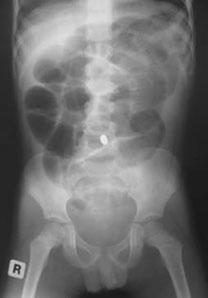
Patient:  A 10 year old female is brought to the ED after swallowing 2 beads (see image).  Based on the findings, what are your concerns and what is the disposition?

Show Answer


An 18-months old presents with classic symptoms of bronchiolitis.  A trial of Albuterol does not show any objective improvement. What are your other options?
- Nebulized epinephrine: 0.9mg/kg for racemic epi or 0.03 mL/kg of the 2.25% solution (diluted in 3mL) - improves oxygen saturation and respiratory rate, but does not affect admission rates
- Hypertonic saline (3%): decreases hospital length of stay and improves clinical scores, possibly by decreasing airway edema and mucus plugging
- Nasal CPAP: improves ventilation in children with bronchiolitis and hypercapnia
- Heliox: decreases respiratory distress, by reducing gaseous flow resistance and improving alveolar ventilation

Interventions that have shown no benefit and are not recommended:
- Anticholinergics
- oral and/or inhaled corticosteroids


Reference:
Joesph, M. Evidence-Based Assessment and Management of Acute Bronchiolitis in the Emergency Department. Pediatric Emergency Medicine Practice 2011; 8(3)


Category: Pediatrics

Title: Nasal foreign body removal

Posted: 12/15/2012 by Mimi Lu, MD (Emailed: 12/21/2012) (Updated: 12/21/2012)
Click here to contact Mimi Lu, MD

Parents bring in their child who placed a bead, seed, or other object up her nose.  What do you do?  Who should you call?

Research suggests that a decades-old home remedy (of sorts) known as the “mother’s kiss” may do the trick for children 1-8 years of age. It’s also much less invasive or frightening than some of the tools and techniques used in emergency departments with a success rate approaching 60%

What Is the “Mother’s Kiss”?

First described in 1965, here’s how the mother’s kiss technique works:

  • The parent or caretaker places their mouth over their child’s mouth while holding the unaffected nostril closed with one finger.
  • The parent or caretaker blows into the child’s mouth.
  • The forceful breath may force the object out (warning: may want to wear protective covering as other things have been known to fly out as well!)


Reference:
Cook S, Burton M, Glasziou P. Efficacy and safety of the "mother's kiss" technique: a systematic review of case reports and case series. CMAJ.2012 Nov 20;184(17):E904-12. doi: 10.1503/cmaj.111864. Epub 2012 Oct 15.


Category: Pediatrics

Title: Fever and neck pain (submitted by Connor Lundy, MD)

Keywords: meningitis, neck pain, retropharyngeal abscess (PubMed Search)

Posted: 11/16/2012 by Mimi Lu, MD
Click here to contact Mimi Lu, MD

Question

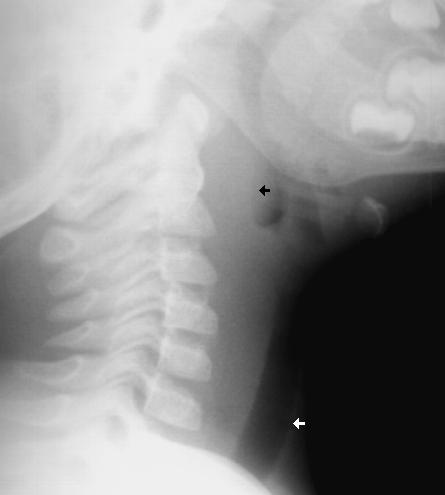
A 1 year old gets sent from their pediatrician’s office for rule out meningitis. They presented with fever for 2 days and neck rigidity. Your LP results are normal. What additional test should you consider?
 

Show Answer


- If child is <6 months think: laryngomalacia and if >6y-3y/o think croup
- The differential of child with stridor <6m:
Ø  laryngomalacia
Ø  vocal cord paralysis
Ø  subglottic stenosis
Ø  vascular ring structures
- Other causes of stridor: tracheitis, epiglottitis, trauma, foreign body, deep neck space infection
- Tips for the treatment of croup:
Ø  Dexmethasone is superior to prednisolone. Start dexmethasone  at 0.15-0.6 mgkg. Typically one time dosing is sufficient. PO/IM forms are considered equivalent.
Ø  A 2011 Cochrane review found no difference in the type of nebulized epinephrine used.
Ø  If regular epinephrine dosing is 0.5 ml/kg of 1:1000. If 2.25% racemic epinephrine, give 0.05 ml/kg.
 
http://www.youtube.com/watch?v=1Enq2BvX9aw&feature=fvwrel
 
References
Donaldson D, et al. Intramuscular versus oral dexamethasone for the treatment of moderate-to-severe croup: a randomized, double-blind trial. Acad Emerg Med. 2003 Jan;10(1):16-21.
Leung AKC, Cho H. Diagnosis of stridor in children. Am Fam Physician. 1999 Nov 15;60(8):2289-2296.
Sparrow A, Geelohoed G.  Prednisolone versus dexamethasone in croup: a randomised equivalence trial. Arch Dis Child. 2006 Jul;91(7):580-3.


The incidence of pediatric syncope is common with 15%-25% of children and adolescents experiencing at least one episode of syncope before adulthood. Incidence peaks between the ages of 15 and 19 years for both sexes.

Although most causes of pediatric syncope are benign, an appropriate evaluation must be performed to exclude rare life-threatening disorders. In contrast to adults, vasodepressor syncope (also known as vasovagal) is the most frequent cause of pediatric syncope (61%–80%).  Cardiac disorders only represent 2% to 6% of pediatric cases but account for 85% of sudden death in children and adolescent athletes.  17% of young athletes with sudden death have a history of syncope.

Key features on history and physical examination for identifying high-risk patients include exercise-related symptoms, a family history of sudden death, a history of cardiac disease, an abnormal cardiac examination, or an abnormal ECG.

Pediatric Dysrhythmias that can cause syncope in children:
- Congenital long QT
- Brugada syndrome
- Catecholaminergic polymorphic VT
- Wolff-Parkinson-White syndrome (WPW)
- Congenital short QT
- Hypertrophic Cardiomyopathy (HCM)
- Arrythmogenic RV dysplasia.
 
 
Reference:
Fischer JW, Cho CS. Pediatric syncope: cases from the emergency department. Emerg Med Clin North Am. 2010 Aug; 28(3):501-16.


Category: Pediatrics

Title: Pediatric intubation (submitted by Danya Khoujah, MBBS)

Keywords: premedication, RSI, ventilator, high flow nasal cannula (PubMed Search)

Posted: 9/21/2012 by Mimi Lu, MD
Click here to contact Mimi Lu, MD

When intubating an infant, a few key points need to be kept in mind:
- Remember that the narrowest point is the cricoid, so even if the ETT passes the cords it might still not pass through the cricoid itself.
- Remember premedication with atropine is recommended in all children less that 1 year old and in those less than 5 years old when using succinylcholine. It is used to prevent reflex bradycardia and high ICP and to decrease secretions. The dose is 0.02 mg/kg IV, with a minimum of 0.1 mg and a max of 0.5 mg. Give it 2 full minutes before the start of intubation.
- Remember that succinylcholine is contraindicated in neuromuscular disease (including an undiagnosed myopathy). A slightly higher dose (2mg/kg) may need to be used in infants (compared to 1-1.5mg/kg in adults and older children).  
- Pressure control mode is preferred over volume control (VC) setting in peds, because VC tends to overestimate how much volume it's delivering, therefore delivering inadequate ventilation.
- Remember your alternatives: High Flow Nasal cannula (HFNC) can be used in infants with respiratory distress to avoid intbation. One study showed that is decreased intubation rates by 68% in respiratory distress due to bronchiolitis
 
References:
1. Santillanes G, Gausche-Hill M. Pediatric Airway Management. Emerg Med Clin N Am 26 (2008) 961–975
2. Bledsoe G H, Schexnayder S M. Pediatric Rapid Sequence Intubation A Review. Ped Emerg Care 20 (2004) 339-344


The mortality from septic shock and severe sepsis ranges between 10-12%.

The PALS algorithm includes 5 points in management.  The first two points are optimally reached within one hour:
1) Recognition of sepsis and vascular access
2) 20ml/kg IVF X 3 within 1 hour or 60ml/kg IVFs within 15 minutes and antibiotic administration
3) Determine if fluid responsive
4) ICU monitoring and/or
5) Vasoactive medications

A recent study at a tertiary care children's hospital retrospectively reviewed 126 patients diagnosed with sepsis. Their findings:

- 37% received 60ml/kg in 60 minutes
- 11% received 60ml/kg in 15 minutes
- 70% received antibiotics in 60 minutes
- In 49% of cases fluids were delivered via IV infusion pump versus manual or pressure bag
- There was a 57% shorter overall hospital stay and 42% shorter ICU stay in patients that received 60ml/kg IVFs within 60 minutes.
- Similarly adherence to the algorithm resulted in decrease hospital stay.
- Liver enzymes, coagulation profiles, and lactic acid levels were obtained in "few" patients.

Conclusions:
Suboptimal fluid resuscitation in sepsis is linked to longer hospital stays. Knowledge of PALS guideline and faster administration of fluid were thought to have been causes of poor adherence.

Additionally, parameters measured in sepsis including lactic acid, coagulation studies, and liver enzymes were not routinely collected. The authors concluded this came from a lack of knowledge of their utility in sepsis.


References:
Paul R, et al. "Adherence to PALS Sepsis Guidelines and Hospital Length of Stay." Pediatrics: 2012 Jul 2 [epub adhead of print].