UMEM Educational Pearls

Title: Scaphoid fractures

Category: Orthopedics

Posted: 8/9/2025 by Brian Corwell, MD (Updated: 12/5/2025)
Click here to contact Brian Corwell, MD

Scaphoid fractures

Make up almost 2/3rds of all carpal fractures

Fractures tend to be localized to 3 anatomic locations

https://cdn.prod.website-files.com/5e97afcf306ae2d7c62acfa5/5ebb8a8dc1ee0554371a19b0_Classification%20copy.png

Waist fractures make up 2/3rds. This may be diagnosed with anatomic snuffbox tenderness

Proximal pole fractures make up approximately 25%. This may be diagnosed with bone tenderness about a fingerbreadth distal to Lister’s tubercle

https://www.orthopaedicsandtraumajournal.co.uk/cms/10.1016/j.mporth.2021.05.001/asset/ae31a4bc-788f-4b41-88c8-a6454ae7645c/main.assets/gr2_lrg.jpg

Distal fractures make up the remaining 10%. This may be diagnosed with tenderness at the volar prominence of the distal wrist crease

https://static.cambridge.org/binary/version/id/urn:cambridge.org:id:binary:54936:20160617100819578-0248:44988fig3_5.png?pub-status=live

Imaging:

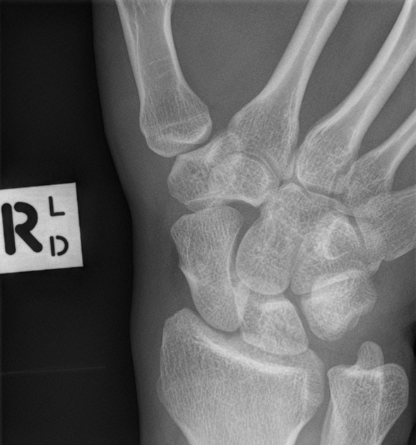
In addition to standard views of the wrist (PA/lateral/oblique) consider adding a scaphoid view. This imaging view is a PA film taken with the wrist in full pronation and ulnar deviation. This allows full visualization of scaphoid in its longitudinal axis. Also, this allows visualization of the area in question without the annoying overlap of adjacent carpal shadows

https://prod-images-static.radiopaedia.org/images/13570123/f9ae97fa07b882fa73ac94b9cf79404a2c05f6974003590e868b6e07f7de83f3_big_gallery.jpeg