This week's visual pearl reviews the structures of the heart when being viewed in a parasternal long-axis view. What do the labels correspond to in the clip below (note: "E" and "F" are valves) and do you see any obvious abnormalities?
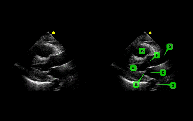
The parasternal long-axis is obtained by scanning to the left (patient's left) of the sternum through the 2nd-5th intercostal space. Click here for a tutorial on the technique.
Answer to Bonus Question: Dilation of the RVOT
Follow me on Twitter (@criticalcarenow) or Google+ (+criticalcarenow)