Category: Visual Diagnosis
Posted: 6/18/2012 by Haney Mallemat, MD
Click here to contact Haney Mallemat, MD
79 year old male with headaches, ataxia, falls, and difficulty urinating. What's the diagnosis?
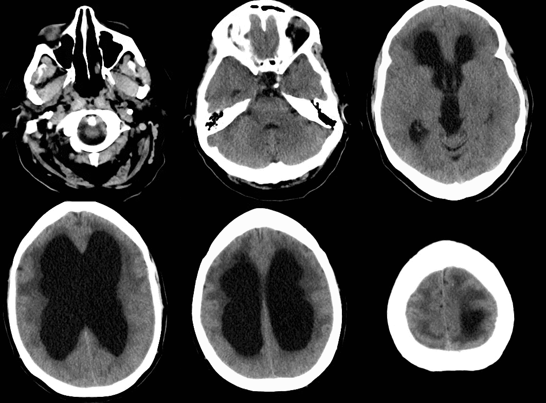
Diagnosis: Ventriculomegaly secondary to Normal Pressure Hydrocephalus
An approach to ventriculomegaly
Ventriculomegaly is due to cerebral atrophy (e.g., Parkinson disease) or increased cerebrospinal fluid (CSF) within the ventricles. Increased CSF is due to:
Congenital causes of ventriculomegaly:
Acquired causes of ventriculomegaly:
Category: Visual Diagnosis
Posted: 6/11/2012 by Haney Mallemat, MD
Click here to contact Haney Mallemat, MD
19 year-old male presents with L ankle pain and obvious deformity after jumping out of a window and landing on his inverted foot. What's the diagnosis?
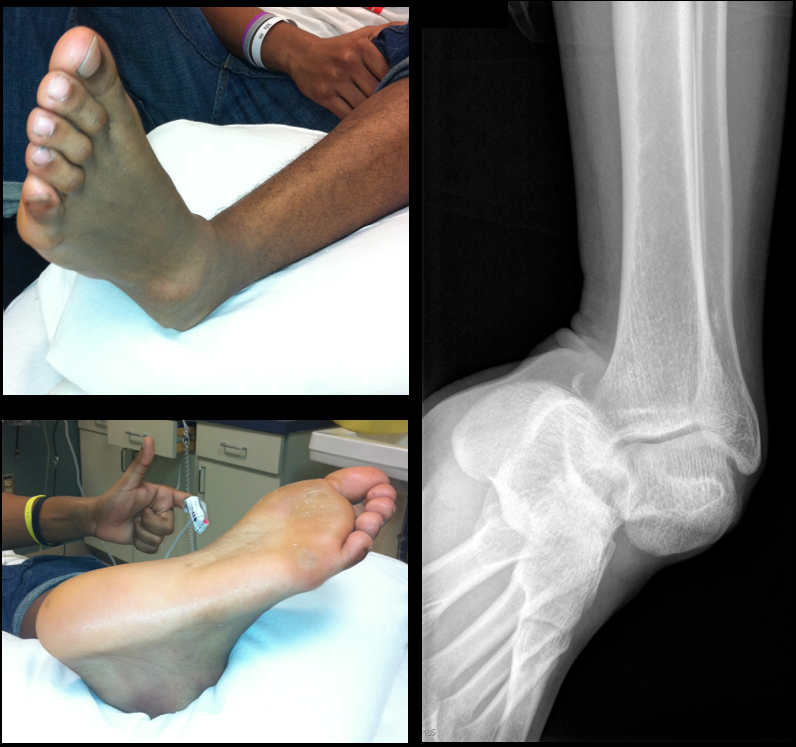
Sub-talar dislocation
Wheeless' Textbook of Orthopedics
Follow me on Twitter (@criticalcarenow) or Google+ (+haney mallemat)
Category: Visual Diagnosis
Posted: 5/28/2012 by Haney Mallemat, MD
Click here to contact Haney Mallemat, MD
Ultrasound is useful during intubation; here is a video explaining how: http://ultrarounds.com/ultrarounds.com/Visual_Pearl_May_28,_2012.html
Today's Bonus Pearl:
EMRA has developed a great antibiotic guide for the iphone (http://itunes.apple.com/us/app/2011-emra-antibiotic-guide/id393020737?mt=8) or android (https://play.google.com/store/apps/developer?id=Emergency+Medicine+Residents'+Association). This app is a bit pricey ($15.99), but is easy to use and well organized. Enjoy!
Chou, H. et al. Tracheal rapid ultrasound exam (T.R.U.E.) for confirming endotracheal tube placement during emergency intubation. Resuscitation. Jun 2011
Werner SL,et al. Pilot study to evaluate the accuracy of ultrasonography in confirming endotracheal tube placement. Ann Emerg Med 2007;49:75–80.
Follow me on Twitter (@criticalcarenow) or Google+ (+haney mallemat)
Category: Visual Diagnosis
Posted: 5/14/2012 by Haney Mallemat, MD
Click here to contact Haney Mallemat, MD
This week's visual pearl is an interesting ultrasound of a psoas abscess submitted by Dr. Sa'ad Lahri. He is an Attending physician in the Emergency Department of the Khayelitsha Hospital in Cape Town, South Africa. The video quality is grainy, but it automatically replays so you can watch it a few times.
http://ultrarounds.com/ultrarounds.com/Visual_Pearl_May_14,_2012.html
Follow me on Twitter (@criticalcarenow) or Google+ (+haney mallemat)
Category: Visual Diagnosis
Posted: 4/29/2012 by Haney Mallemat, MD
(Updated: 4/30/2012)
Click here to contact Haney Mallemat, MD
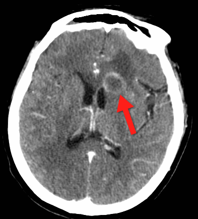
68 yo man presents with new-onset seizures; his CT is shown below. What is your differential diagnosis?
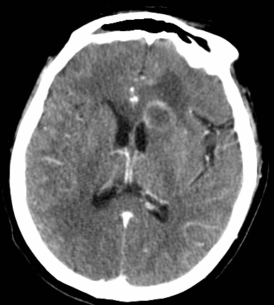
Cerebral Ring-Enhancing Lesions
Neoplasm
Infectious
Neurologic
Bonus pearl: Do you like Emergency Ultrasound and want a quick review before you scan your next patient? Well, check out the "One-minute Ultrasound App". It's provides a quick review for many essential ultrasound studies...and yes, it's FREE for both iphone and android.
Iphone: http://itunes.apple.com/us/app/one-minute-ultrasound/id512301845?mt=8&ls=1
Garg, R., Sinha, M. Multiple ring-enhancing lesions of the brain. J Postgrad Med. 2010 Oct-Dec; 56(4):307-16
Follow me on Twitter (@criticalcarenow) or Google+ (+haney mallemat)
Category: Visual Diagnosis
Posted: 4/15/2012 by Haney Mallemat, MD
(Updated: 4/16/2012)
Click here to contact Haney Mallemat, MD
67 yo male presents with burning substernal chest pain; worse with meals and when supine. What's the diagnosis?
Answer: Hiatal hernia
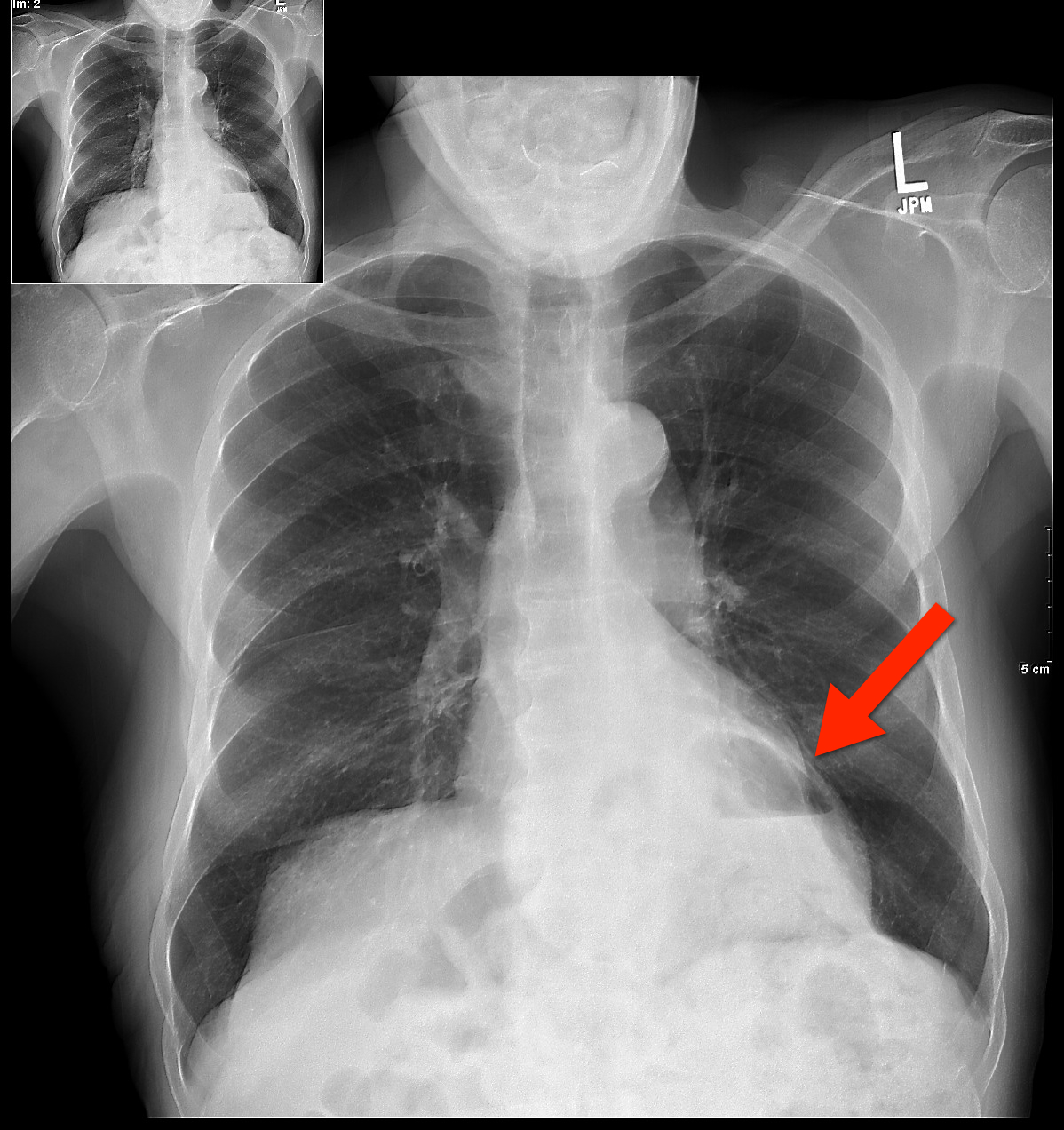
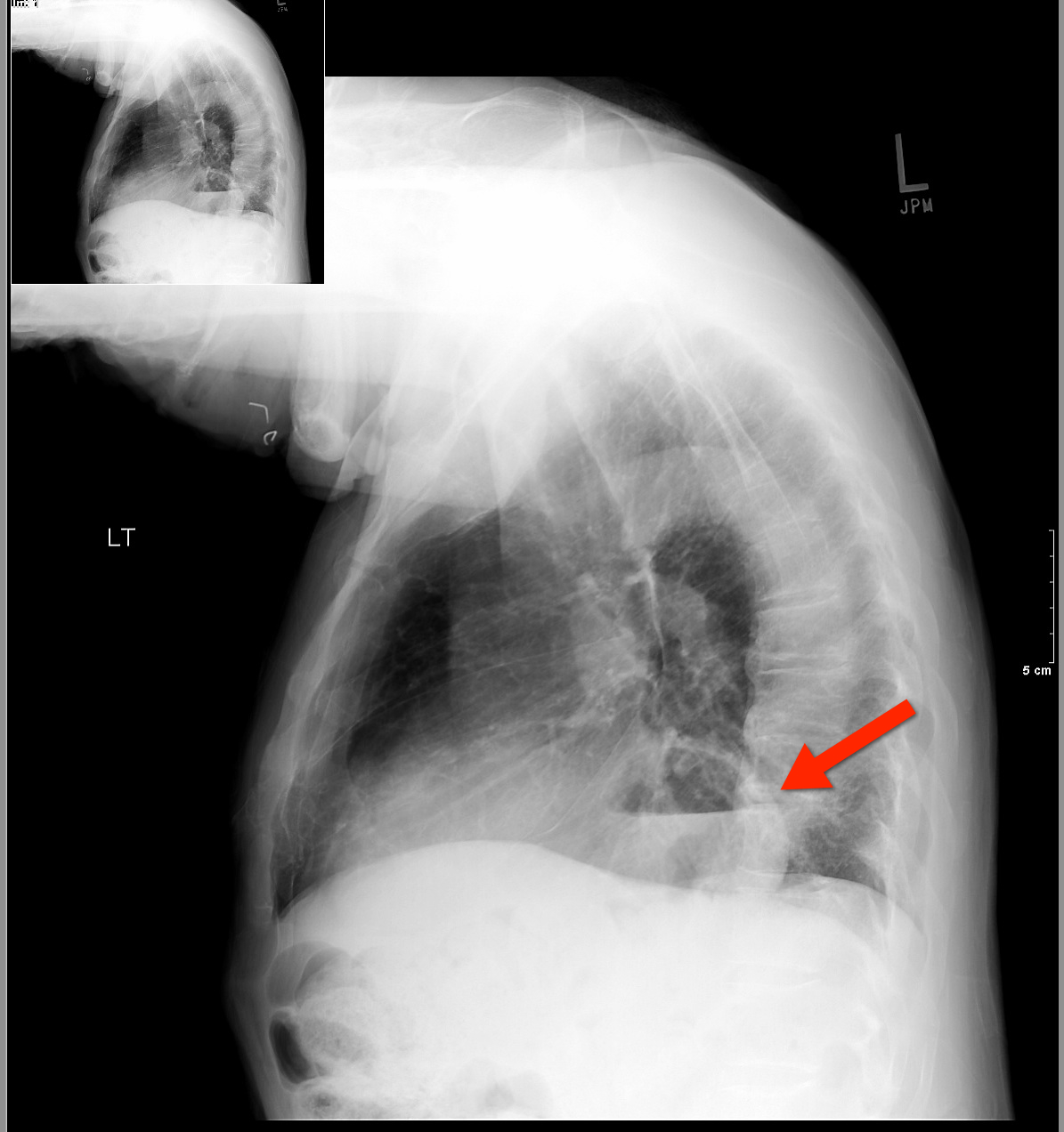
The differential diagnosis for circumscribed air-fluid levels on chest X-ray includes:
Bonus Pearl
Do you like listening to pre-recorded lectures, especially when they’re free? Then check out Free Emergency Medicine Talks (http://freeemergencytalks.net) where you can listen to millions (ok, more like 1,384) of free lectures recorded at major conferences around the world.
Follow me on Twitter (@criticalcarenow) or Google+ (+haney mallemat)
Category: Visual Diagnosis
Posted: 3/25/2012 by Haney Mallemat, MD
(Updated: 3/26/2012)
Click here to contact Haney Mallemat, MD
26 year old male with pain when he extends his 4th finger as well as swelling of that digit. Diagnosis?
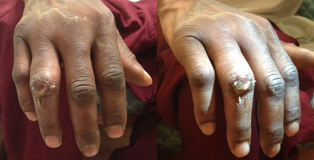
Answer: Infectious Flexor Tenosynovitis
Infectious Flexor Tenosynovitis
- Closed space infection of the flexor tendon sheath; an orthopedic emergency
- Typically an infection with skin flora secondary to penetrating trauma
- Diagnosed by Kanavel's Signs:
- Finger held in slight flexion
- Fusiform swelling
- Pain on passive extension
- Pain while palpating the tendon sheath
- If early and not severe a course of IV antibiotics (covering skin flora) may be tried, however, surgical intervention is often necessary; early consultation with a hand surgeon is highly recommended
Follow me on Twitter (@criticalcarenow) or Google+ (+haney mallemat)
Category: Visual Diagnosis
Posted: 3/12/2012 by Haney Mallemat, MD
(Updated: 8/12/2014)
Click here to contact Haney Mallemat, MD
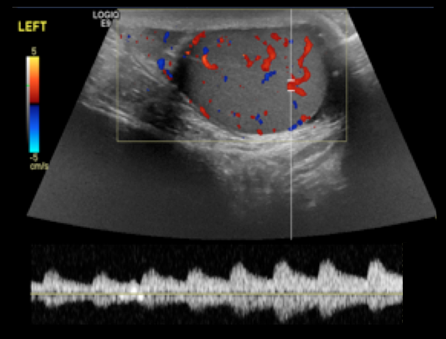
14 year-old male presents with right-sided testicular pain. What's the diagnosis?
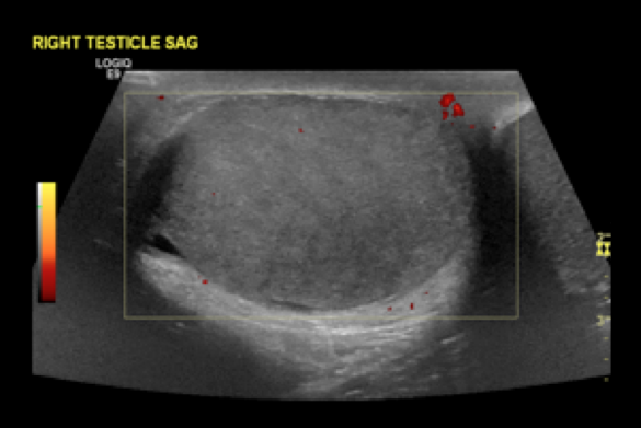
Answer: Testicular torsion
References
Shan Yin, MD, MPH, Jennifer L. Trainor, MD, Diagnosis and Management of Testicular Torsion, Torsion of the Appendix Testis, and Epididymitis, Clin Ped Emerg Med, 10:38-44, 2009
Follow me on Twitter (@criticalcarenow) or Google+ (+haney mallemat)
Category: Visual Diagnosis
Posted: 2/27/2012 by Haney Mallemat, MD
(Updated: 8/28/2014)
Click here to contact Haney Mallemat, MD
24 year-old male presents following fall from a scaffolding and complains of wrist pain. Diagnosis?
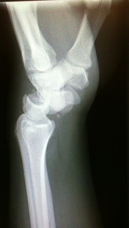
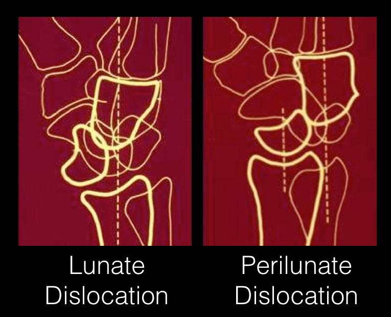
Answer: Perilunate dislocation
Perilunate dislocations usually occur following high-energy trauma (e.g., fall from a height); the mechanism is usually wrist hyperextension, ulnar deviation, and carpal supination.
Tenderness is palpated along the dorsum of the wrist; specifically distal to the lister tubercle along the scapholunate ligament; injuries may be associated with scaphoid fracture
Paresthesias may also occur along the median nerve distribution with up to 25% of cases developing carpal tunnel syndrome.
Treatment options include closed reduction and casting or open reduction and ligamentous repair with internal fixation.
The figure below illustrates the differences between lunate and perilunate dislocations.
References
Lunate and Perilunate Dislocations. Kannikeswaran, N & Sethuraman, U.
http://www.radiologyassistant.nl/en/42a29ec06b9e8 (diagram)
Follow me on Twitter (@criticalcarenow) or Google+ (+haney mallemat)
Category: Visual Diagnosis
Posted: 2/13/2012 by Haney Mallemat, MD
(Updated: 8/28/2014)
Click here to contact Haney Mallemat, MD
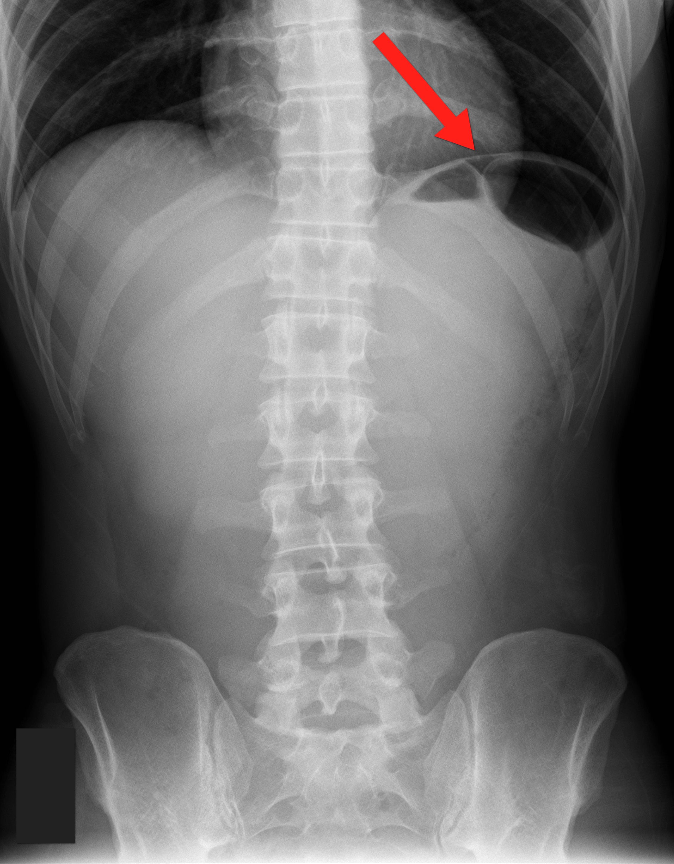
35 year old male with sudden onset of abdominal pain. Diagnosis?
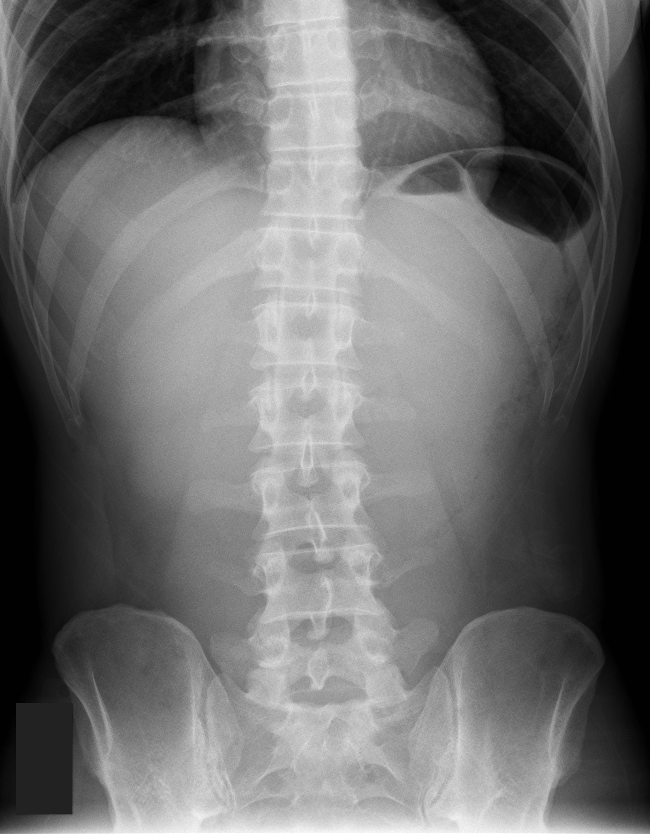
Answer: Pneumoperitoneum
Follow me on Twitter (@criticalcarenow) or Google+ (+haney mallemat)
Category: Visual Diagnosis
Posted: 2/5/2012 by Haney Mallemat, MD
(Updated: 8/28/2014)
Click here to contact Haney Mallemat, MD
28 y.o. male felt his left knee "pop" after landing from a jump while playing basketball. Knee exam revealed limited knee extension. X-ray is shown. Diagnosis?
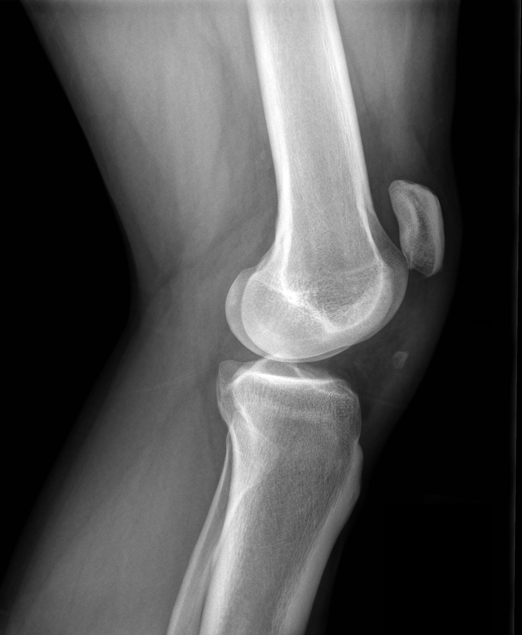
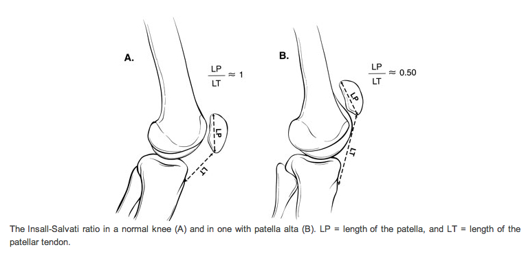
Answer: Patella Alta secondary to Patellar tendon rupture
References
Follow me on Twitter (@criticalcarenow) or Google+ (+haney mallemat)
Category: Visual Diagnosis
Posted: 1/22/2012 by Haney Mallemat, MD
(Updated: 1/23/2012)
Click here to contact Haney Mallemat, MD
20 year old female complains of “itchy” rash to her foot x 1 week and recently the rash has spread to her other other foot and both hands (shown below). No past medical history, no fever or chills, no mucus membranes involvement, no new medications, no tick bites, no travel. She is also 16 weeks pregnant. What’s the diagnosis?
Answer: Secondary syphilis
Follow me on Twitter (@criticalcarenow) or Google+ (+haney mallemat)
Category: Visual Diagnosis
Posted: 1/9/2012 by Haney Mallemat, MD
(Updated: 8/28/2014)
Click here to contact Haney Mallemat, MD
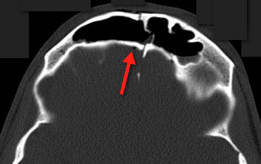
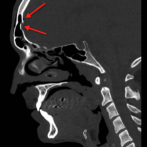
23 year-old male fell off porch while intoxicated. The head CT is shown below. Diagnosis?
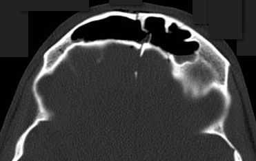
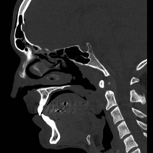
Answer: Frontal sinus fracture (inner and outer table) with pneumocephalus.
A few quick pearls when managing skull fractures:
Medical management:
Surgical management, if:
Follow me on Twitter (@criticalcarenow) or Google+ (+haney mallemat)
Category: Visual Diagnosis
Posted: 12/26/2011 by Haney Mallemat, MD
Click here to contact Haney Mallemat, MD
64 year old male with emphysema and stage 4 lung cancer presents in respiratory distress. What's the diagnosis?
Answer: Pneumothorax (left chest) and bullous disease (right chest).
In questionable cases like this, bedside ultrasound can help distinguish between bullae and pneumothorax. Bullae should have a positive lung sliding sign whereas pneumothorax does not.
See the referenced case report for more information:
Simon, B. et al. Two cases where bedside ultrasound was able to distinguish pulmonary bleb from pneumothorax. J Emerg Med. 2005 Aug;29(2):201-5.
Follow me on Twitter (@criticalcarenow) or Google+ (+haney mallemat)
Category: Visual Diagnosis
Posted: 12/11/2011 by Haney Mallemat, MD
(Updated: 8/28/2014)
Click here to contact Haney Mallemat, MD
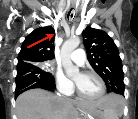
60 year old male with 6 months of weight loss and recent epistaxis. Diagnosis?
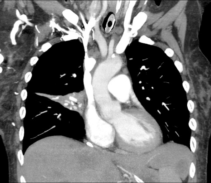
Answer: Partial obstruction of the Superior vena cava (SVC)
SVC Syndrome
SVC syndrome is caused by compression or obstruction of the superior vena cava blocking anterograde flow.
Most cases are secondary to extrinsic compression by malignancy. Other causes are secondary to thrombosis and internal obstruction (e.g., central venous catheter placement).
Symptoms present sub-acutely, worsen with bending over, and are secondary to increased venous pressure in the head and neck (e.g., epistaxis, headache, tinnitus, conjunctival injection, neck swelling, etc.).
Treatment focuses on reversing the underlying cause (e.g., radiation or chemotherapy if due a sensitive tumor) and treatment of symptoms:
Rice TW, Rodriguez RM, Light RW. The superior vena cava syndrome: clinical characteristics and evolving etiology. Medicine (Baltimore). Jan 2006;85(1):37-42.
Nunnelee JD. Superior vena cava syndrome. J Vasc Nurs. Mar 2007;25(1):2-5; quiz 6.
Follow me on Twitter (@criticalcarenow) or Google+ (+haney mallemat)
Category: Visual Diagnosis
Posted: 11/27/2011 by Haney Mallemat, MD
(Updated: 11/28/2011)
Click here to contact Haney Mallemat, MD
9 year-old boy with sudden onset of unilateral facial swelling. What’s the diagnosis?

Answer: Acute Parotitis
Bonus Trivia: U.S. President Garfield died from parotitis after becoming dehydrated following abdominal surgery
Shelly J. McQuone MD, Acute Viral and Bacterial Infections of the Salivary Glands, Otolaryngologic Clinics of North America, Volume 32, Issue 5 (October 1999)
Follow me on Twitter (@criticalcarenow) or Google+ (+haney mallemat)
Category: Visual Diagnosis
Posted: 10/30/2011 by Haney Mallemat, MD
(Updated: 10/31/2011)
Click here to contact Haney Mallemat, MD
72 year-old man, one-week post right fem-pop bypass presents with painful blue and black toe. Diagnosis?
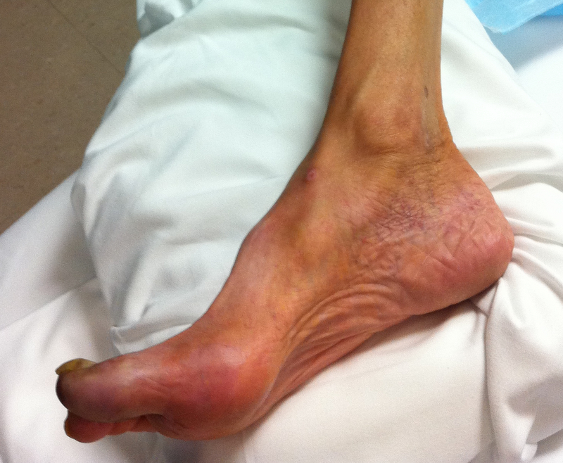
Answer: Blue-Toe Syndrome
Hirschman, J. et al. Blue (or purple) toe syndrome. J Am Acad Dermatol.2009 Jan;60(1):1-20
O'Keeffe S, et al. Blue toe syndrome: Causes and management.Arch Intern Med. 1992 Nov;152(11):2197-202.
Follow me on Twitter (@criticalcarenow) or Google+ (+haney mallemat)
Category: Visual Diagnosis
Posted: 10/17/2011 by Haney Mallemat, MD
Click here to contact Haney Mallemat, MD
5 year-old male with developmental delay presents with intractable non-bloody and non-bilious vomiting over 10 days; bowel movements are normal. Four weeks ago he was placed in a hip-spica cast following a motor vehicle crash. Abdominal x-ray is below. Diagnosis?
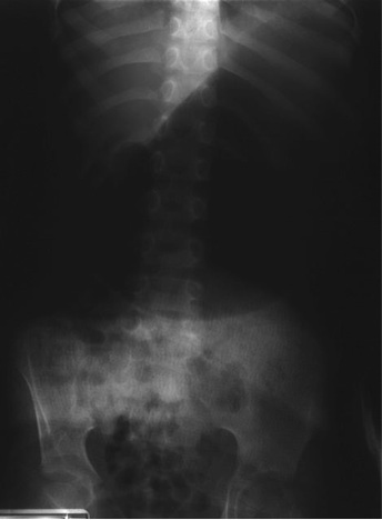
Answer: CAST syndrome (also known as Superior Mesenteric Artery Syndrome)
Wheeless Textbook of Orthopedics. Updated August 29,2011
Lichenstein, R. Radiology Cases in Pediatric Emergency Medicine, Volume 5, Number 16
Follow me on Twitter (@criticalcarenow) or Google+ (+haney mallemat)
Category: Visual Diagnosis
Posted: 10/3/2011 by Haney Mallemat, MD
Click here to contact Haney Mallemat, MD
Question: 50-year-old diabetic female s/p foot burn several weeks ago, now presenting with pain and discharge from a poorly healing wound. Diagnosis?
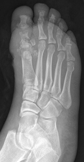
Answer: Osteomyelitis
Osteomyelitis
· Acute or chronic bone infection
· Risk factors: Immunosuppression (diabetes, chronic steroid use, AIDS, and sickle-cell dz.)
· Secondary to direct trauma, contiguous spread from local infection, or hematogenous spread (in children)
· Common bacteria: S. Aureus, Pseudomonas, Salmonellae (classically in Sickle cell dz.)
· X-ray (limited sensitivity):
- 3-5 days post-infection: Soft-tissue swelling
- 14-21 days: Some patients demonstrate bony changes (e.g., periosteal elevation, bone lucencies, etc.)
- >28 days: >90% with Xray findings
· MRI is the imaging gold standard
· Two of the following needed for diagnosis:
- Purulent aspiration
- Positive blood or tissue culture
- Positive imaging
- Tenderness + erythema / edema
· Antibiotic coverage based on culture results. When immediate empiric therapy required (sepsis), cover most likely pathogen plus MRSA.
References
Carek PJ, Dickerson LM, Sack JL. Diagnosis and management of osteomyelitis. Am Fam Physician. 2001 Jun 15;63(12):2413-20.
Pruthi S, Thapa MM. Infectious and inflammatory disorders. Radiol Clin North Am. Nov 2009;47(6):911-26.
Zink BJ, Raukar NP. Bone and Joint Infections. In: Marx JA, Hockberger RS, Walls RM, Adams JG, Barsan WG, Biros MH, Danzl DF, Gausche-Hill M, Ling LJ, Newton EJ, eds. 7th ed. Emergency Medicine: Concepts and Clinical Practice.Volume 2. Philadelphia, PA: Mosby; 2010:1821-1830.
Follow me on Twitter (@criticalcarenow) or Google+ (+haney mallemat)
Category: Visual Diagnosis
Posted: 9/19/2011 by Haney Mallemat, MD
(Updated: 8/28/2014)
Click here to contact Haney Mallemat, MD
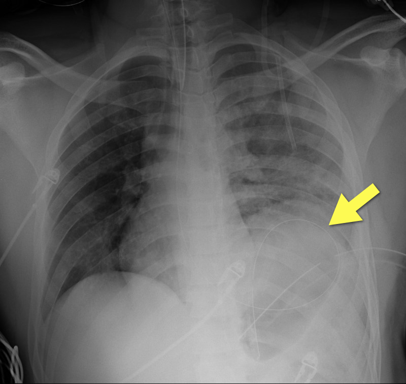
19 year-old male s/p high-speed MVC with hypotension and diminished breath sounds on left. Diagnosis?
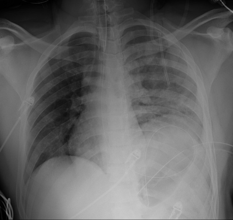
Answer: Diaphragmatic rupture (Note position of NG tube on image below).
·Uncommon; less than 1% of all traumatic injuries
·Diagnosis may be obvious on CXR (as in example) or subtle (e.g., diaphragmatic elevation, basilar atelectasis, etc.)
·Requires a high-index of suspicion because delayed diagnosis increases the risk of abdominal organ herniation or strangulation.
·Mortality depends on the mechanism and presence of associated injuries; mortality is highest for blunt injuries.
Bhavnagri S, et al. When and how to image a suspected broken rib. Cleveland Clinic J Med. 2009;76(5):309.
Williams M, et al. Predictors of mortality in patients with traumatic diaphragmatic rupture and associated thoracic and/or abdominal injuries. Am Surg. 2004;70(2):157.
National Trauma Data Base. American College of Surgeons 2000-2004.
Follow me on Twitter (@criticalcarenow) or Google+ (+haney mallemat)
