Category: Visual Diagnosis
Posted: 9/29/2013 by Haney Mallemat, MD
(Updated: 9/30/2013)
Click here to contact Haney Mallemat, MD
65 year-old diabetic patient presents with abdominal pain. What's the abnormality on Xray?
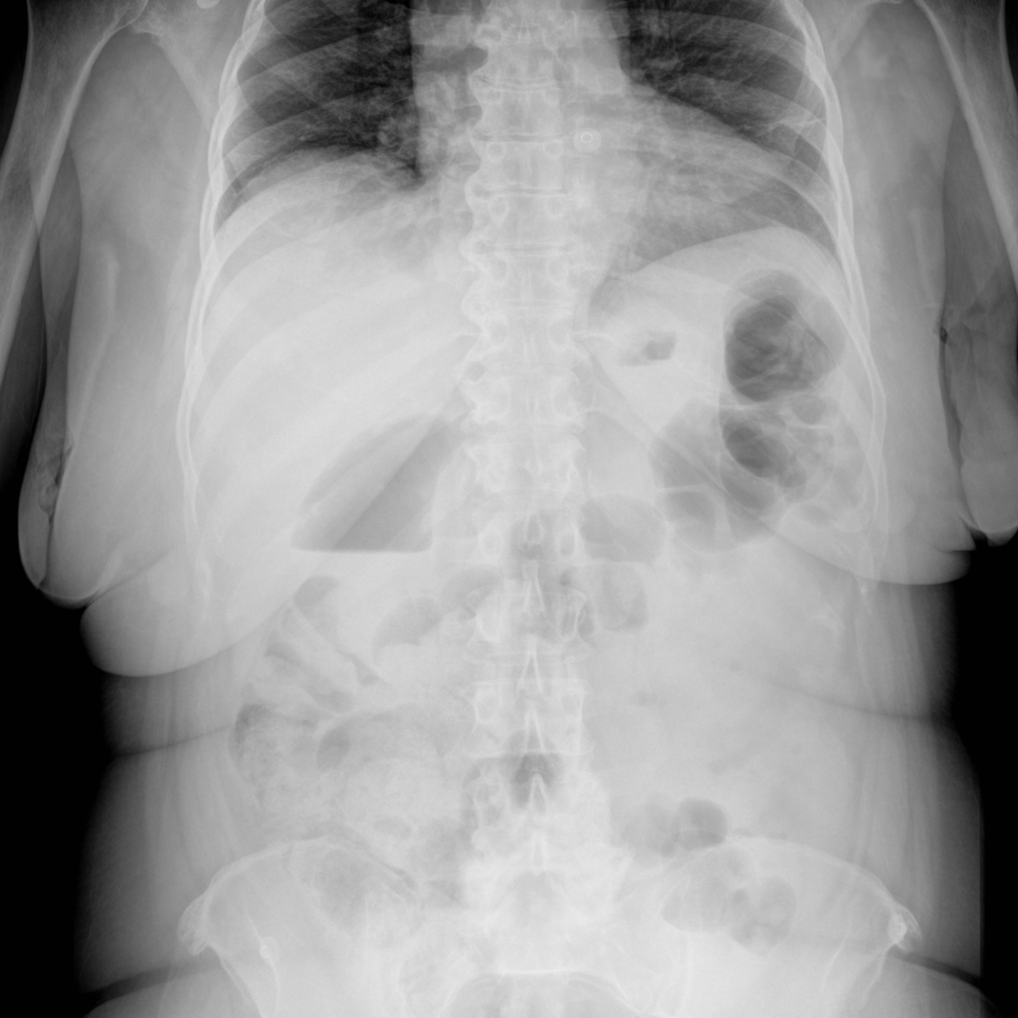
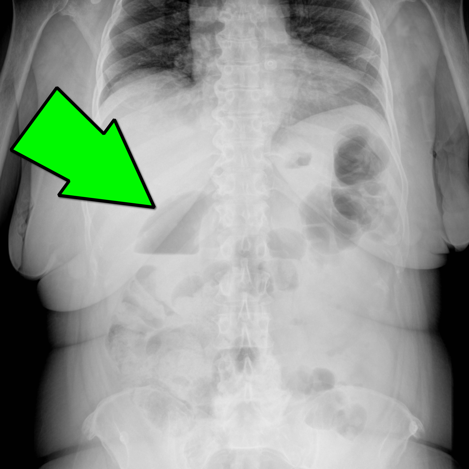
Emphysematous Cholecystitis
Emphysematous Cholecystitis
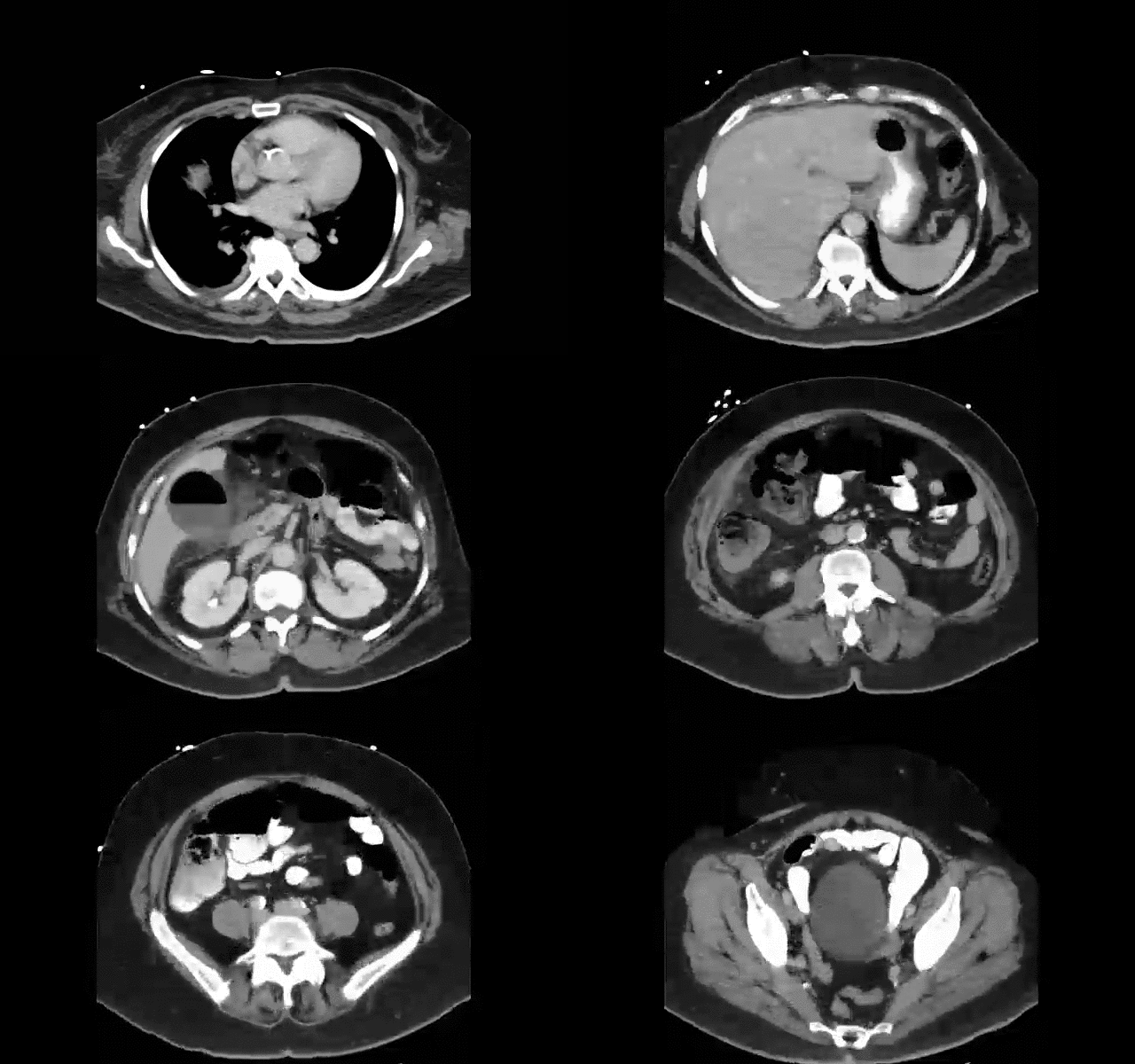
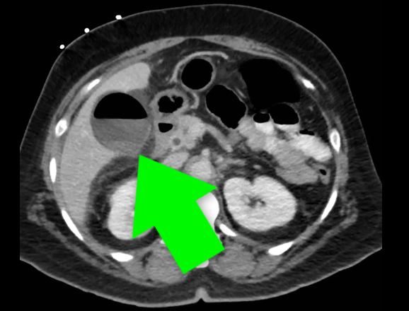
Carrascosa MF, et al. Emphysematous Cholecystitis. CMAJ.10 Jan 2012; 184(1): E81
Category: Visual Diagnosis
Posted: 9/23/2013 by Haney Mallemat, MD
Click here to contact Haney Mallemat, MD
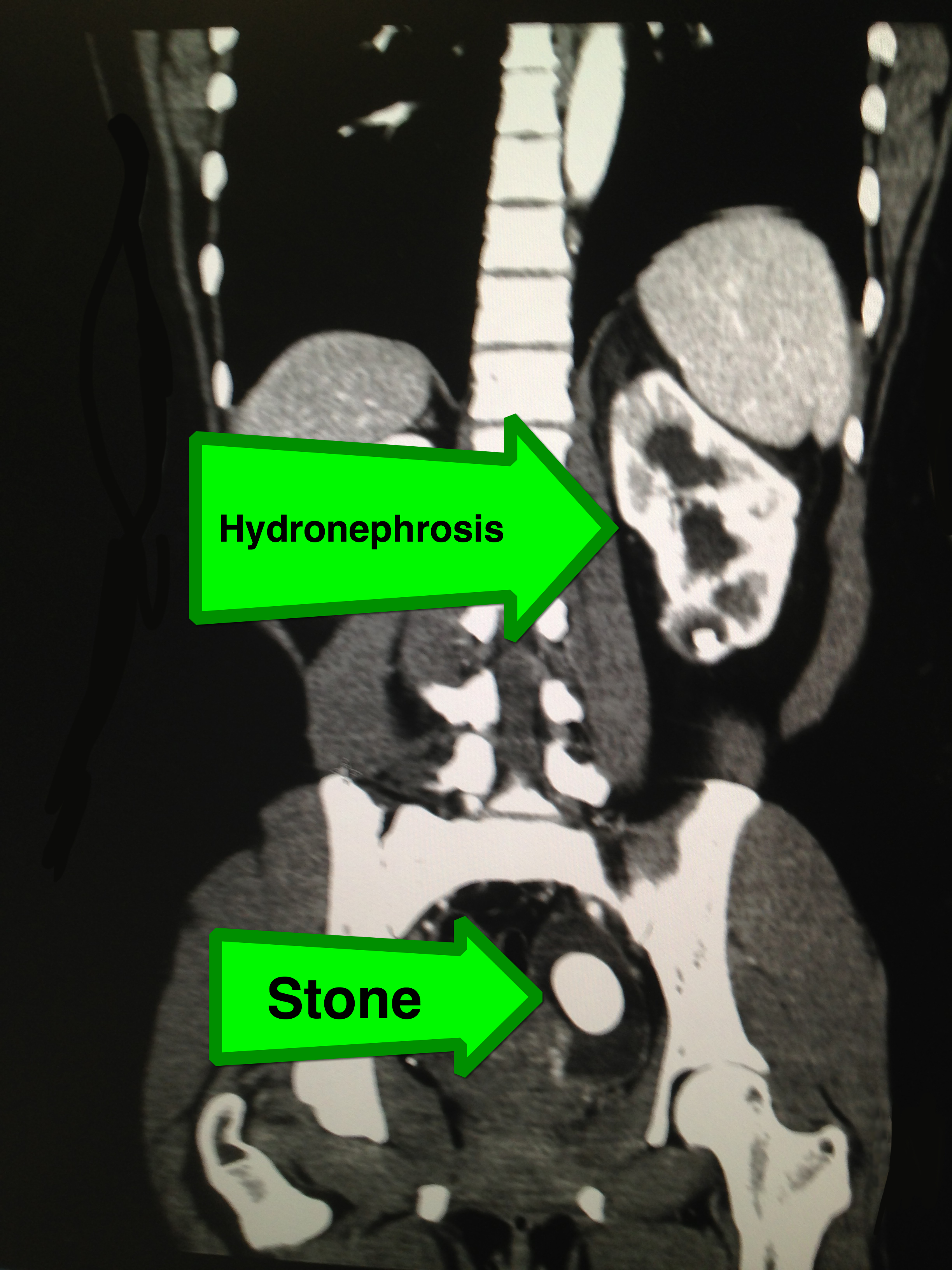
27 year-old female with no past medical history presents with sudden onset of left lower quadrant pain. What's the diagnosis?
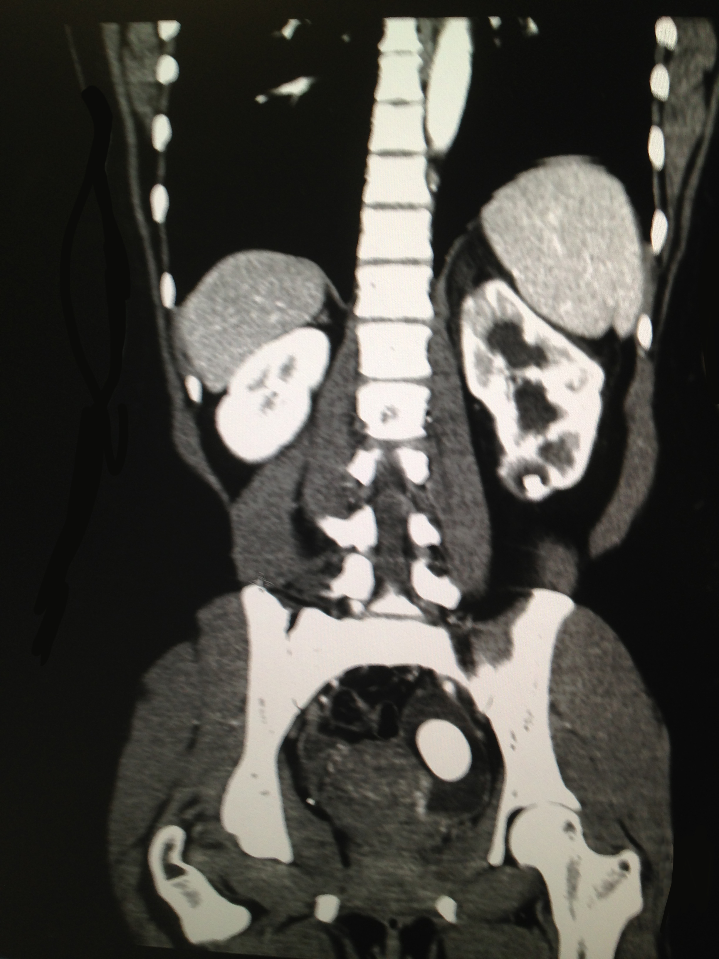
Large left-sided ureterolithiasis with hydronephrosis
Category: Visual Diagnosis
Posted: 9/16/2013 by Haney Mallemat, MD
Click here to contact Haney Mallemat, MD
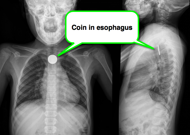
8 year-old girl presents with dysphagia and drooling, Xray is shown. What’s the diagnosis (and where is it located)?
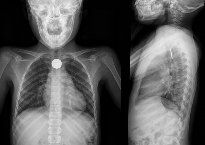
A coin located in the esophagus at the level of the cricopharyneus muscle
Foreign body (FB) pearls
Kenton, Foreign Bodies in the Gastrointestinal Tract and Anorectal Emergencies, Emerg Med Clin N Am 29 (2011) 369–400
Category: Visual Diagnosis
Posted: 9/8/2013 by Haney Mallemat, MD
(Updated: 9/9/2013)
Click here to contact Haney Mallemat, MD
This week's case is challenging, but very interesting...
An elderly patient presents with a history of significant weight loss and chronic constipation; abdominal Xray is below. What's the diagnosis? (Hint: why is the right kidney and psoas muscle so well defined?)
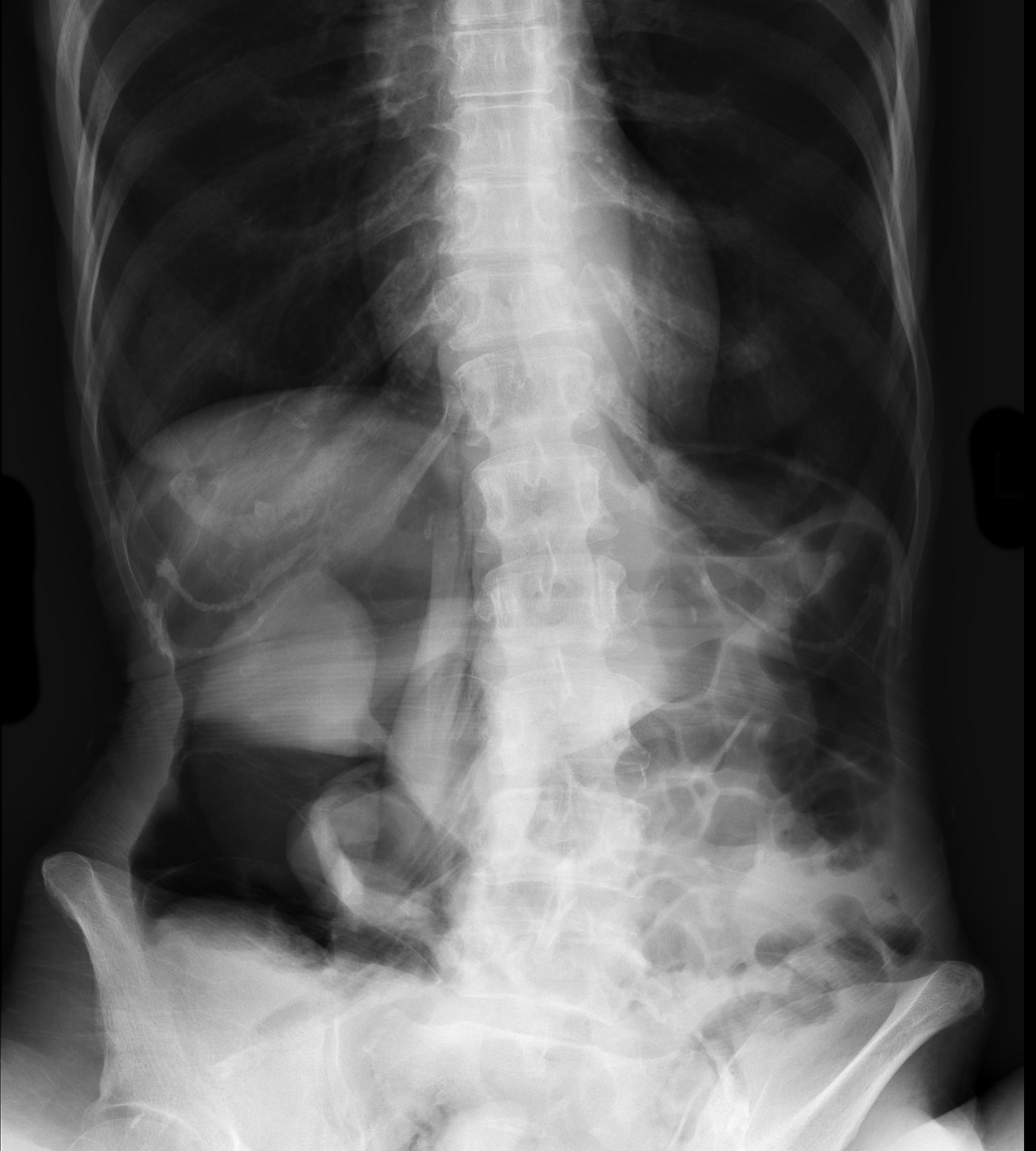
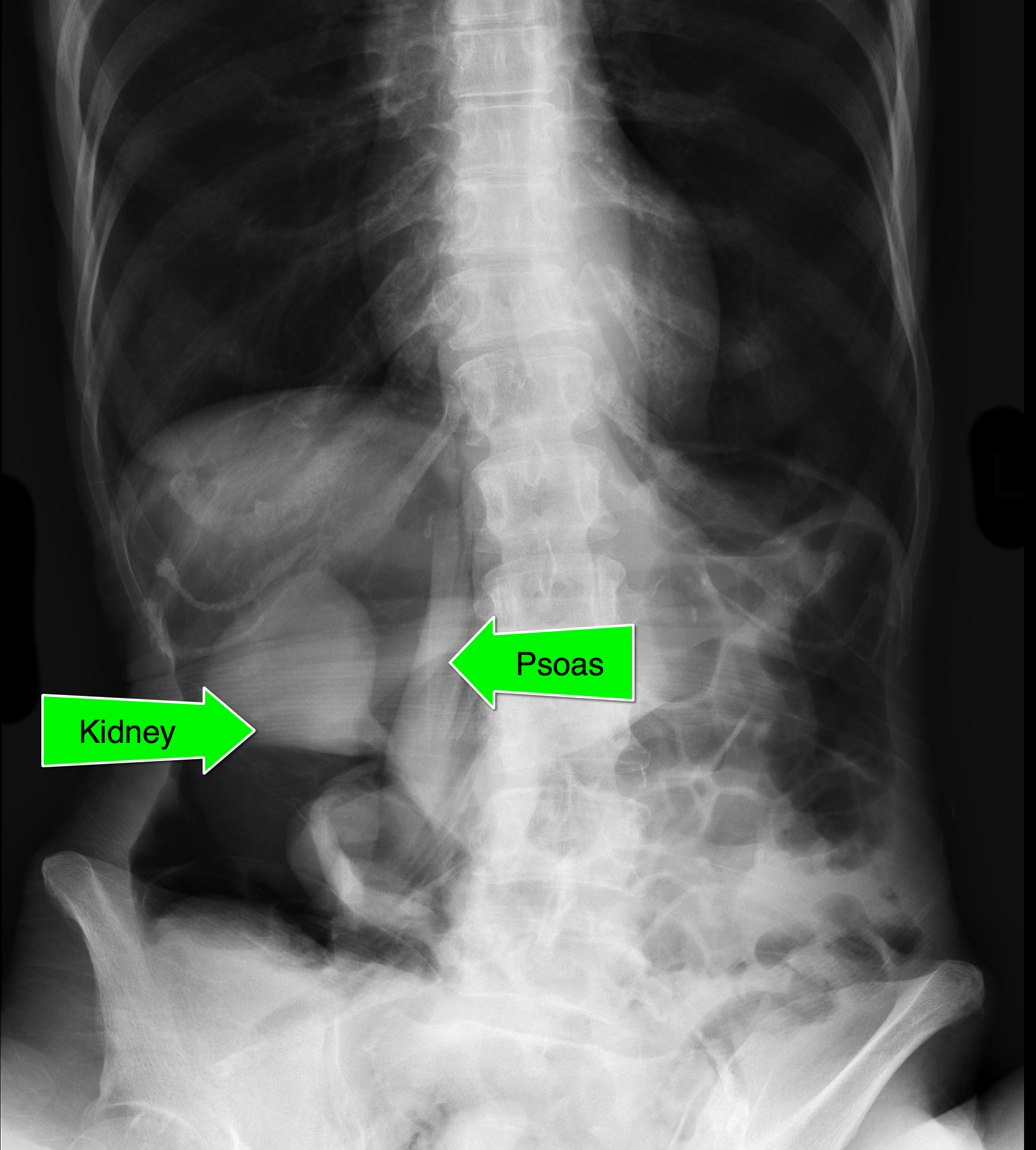
Follow me on Twitter (@criticalcarenow) or Google+ (+criticalcarenow)
Category: Critical Care
Posted: 9/3/2013 by Haney Mallemat, MD
Click here to contact Haney Mallemat, MD
UEDVT comprise 10% of all DVTs (majority are lower extremity), but incidence of UEDVT is rising; UEDVTs are categorized into distal (veins distal to axillary vein) or proximal (from superior vena cava to axillary vein)
Compared to lower extremity DVT, UEDVTs have lower:
75% of UEDVT are secondary (indwelling catheters, pacemakers, malignancy, etc.) and 25% are primary in nature; #1 primary cause of UEDVT is Paget – Schroetter disease
Up to 25% of patients with primary UEDVTs are eventually found to have an underlying malignancy; patients with idiopathic UEDVT should be referred for cancer workup
Treatment includes removal of the catheter (if no longer needed) and:
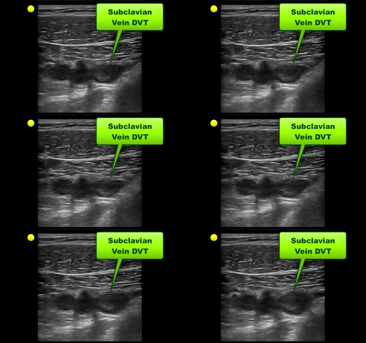
Follow me on Twitter (@criticalcarenow) or Google+ (+criticalcarenow)
Category: Visual Diagnosis
Posted: 9/2/2013 by Haney Mallemat, MD
Click here to contact Haney Mallemat, MD
Elderly male presents with headache, confusion, and trouble with gait. What's in your differential diagnosis?
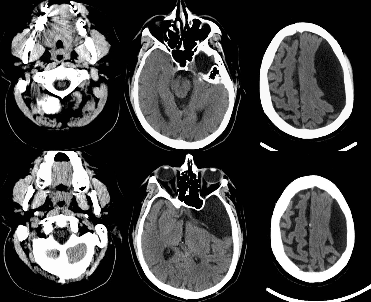
Based on the CT scan shown, the differential here includes epidermoid and arachnoid cyst
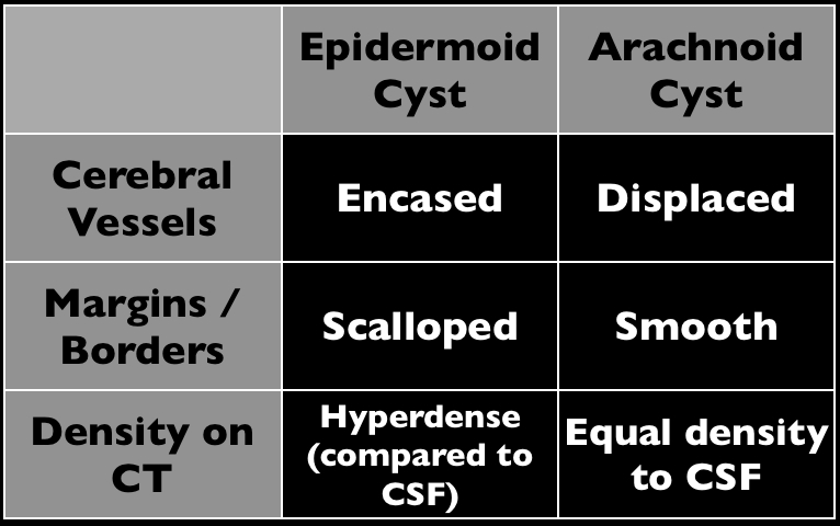
Arachnoid cysts (AC) occur within the cerebrospinal axis and do not communicate with the ventricular system. Most occur in the middle cranial fossa and are typically benign; continuing cerebrospinal fluid
The majority of AC occurs from abnormalities in development, but a small portion occurs secondary to post-surgical adhesions or in association with cancer.
MRI is the test of choice to help define the extent of the cyst as well as determine alternative diagnoses.
Treatment is variable with some experts stating that only symptomatic ACs should be treated with others recommending removal to avoid future complications.
The patient in the stem presented with symptoms secondary to complications from the AC.
Follow me on Twitter (@criticalcarenow) or Google+ (+criticalcarenow)
Category: Visual Diagnosis
Posted: 8/25/2013 by Haney Mallemat, MD
(Updated: 8/26/2013)
Click here to contact Haney Mallemat, MD
23 year-old patient presents with a rash on his palms and soles. He also states that he had a something strange on his genitals several weeks before. What's the diagnosis and what’s the treatment (including dosing) for this disease?
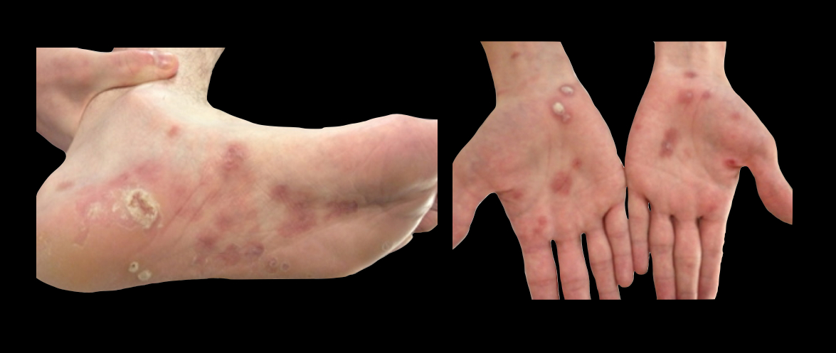
Centers for Disease Control and Prevention, National Center for HIV/AIDS, Viral Hepatitis, STD, and TB Prevention, Division of STD Prevention. 2010 Treatment Update.
http://www.cdc.gov/std/syphilis/treatment.htm
Follow me on Twitter @criticalcarenow or Google+ (+criticalcarenow)
Category: Visual Diagnosis
Posted: 8/19/2013 by Haney Mallemat, MD
Click here to contact Haney Mallemat, MD
Which echocardiographic view of the heart is this and can you name all 6 segments of the left ventricle? (Hint: A = Anteroseptal wall)
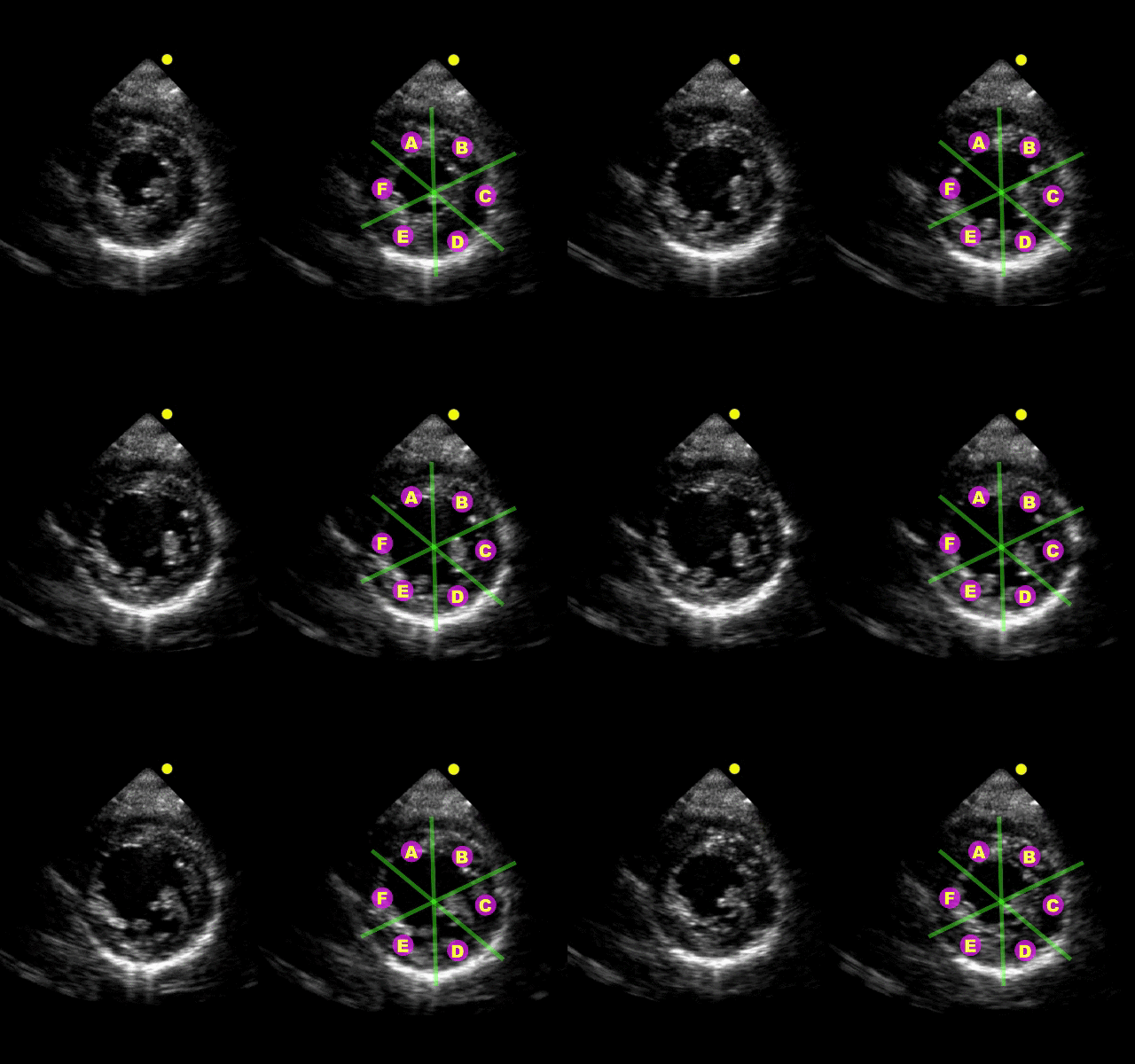
Parasternal short-axis view at the level of the papillary muscles

Category: Visual Diagnosis
Posted: 8/10/2013 by Haney Mallemat, MD
(Updated: 8/12/2013)
Click here to contact Haney Mallemat, MD
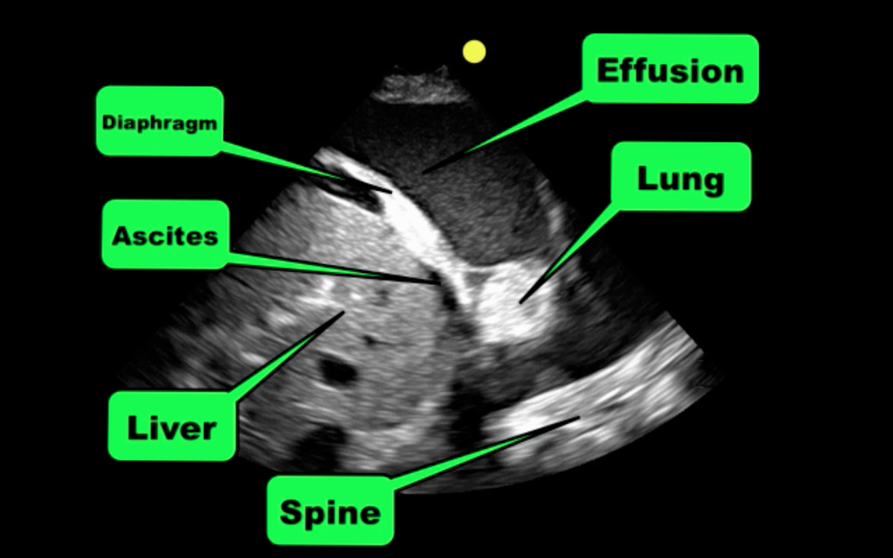
Patient with liver disease presents with dyspnea, fever, and the following ultrasound? What's the diagnosis? (Hint: there are two)?
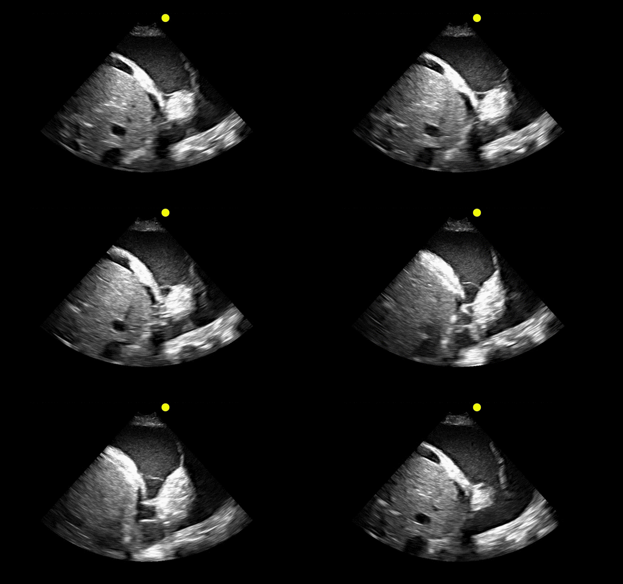
Answer: Complex pleural effusion and ascites
The take away point from this case is to always place the diaphragm in the center of the screen in order to distinguish peritoneal from thoracic fluid. Fluid in both compartments will sometimes be present (as in this case).
Complex pleural effusions
Follow me on Twitter (@criticalcarenow) or Google+(+criticalcarenow)
Category: Visual Diagnosis
Posted: 8/5/2013 by Haney Mallemat, MD
Click here to contact Haney Mallemat, MD
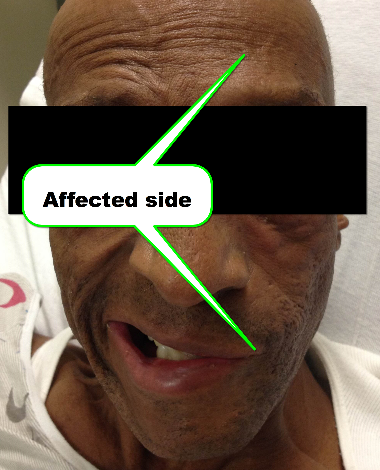
45 year-old man presents after he cannot close his left eye. In the photo below, he is trying to simultaneously raise his forehead and smile. Of note, he was also started on doxycycline recently for Lyme disease. What two medications should he receive?

Bell Palsy
Follow me on Twitter (@criticalcarenow) or Google+ (+criticalcarenow)
Category: Critical Care
Posted: 7/30/2013 by Haney Mallemat, MD
Click here to contact Haney Mallemat, MD
Elderly patient who originally presented for severe pancreatitis now intubated for worsening hypoxemia. CXR is shown below, what's the diagnosis?
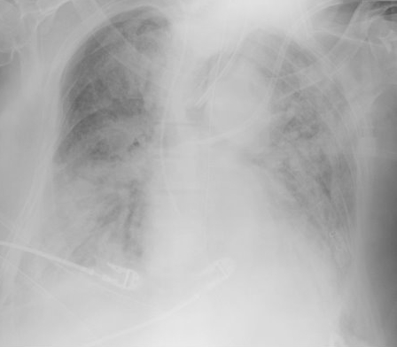
Acute Respiratory Distress Syndrome (ARDS)
Acute Respiratory Distress Syndrome (ARDS) is defined as hypoxemia secondary to increased pulmonary capillary permeability and non-hydrostatic (i.e., non-cardiogenic) leakage of fluid into the interstitial lung tissue and alveoli. Lung radiographs diffuse and symmetric infiltrates (see below)
ARDS may occur secondary to a primary (or pulmonary) insult (e.g., aspiration, pneumonia) or secondary (or systemic) insult (e.g., pancreatitis, trauma, etc.)
The newest classification system for ARDS no longer includes the previously known category of acute lung injury; there are three categories of ARDS determined by the PaO2 (on ABG) divided by administered FiO2 (as a fraction of 100%):
A number of interventions have been demonstrated to improve outcomes for patients with ARDS:
Follow me on Twitter (@criticalcarenow) or on Google+ (+criticalcarenow)
Category: Visual Diagnosis
Posted: 7/29/2013 by Haney Mallemat, MD
Click here to contact Haney Mallemat, MD
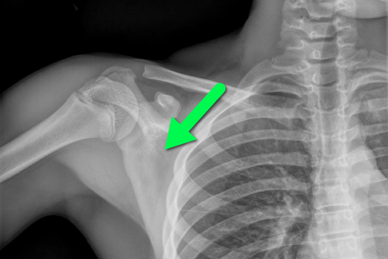
13 year-old female fell on right shoulder while catching a rebound during a basketball game. The patient is holding her arm in adduction and has exquisite scapular tenderness on exam. What’s the next step in management? …oh, and what’s the diagnosis?
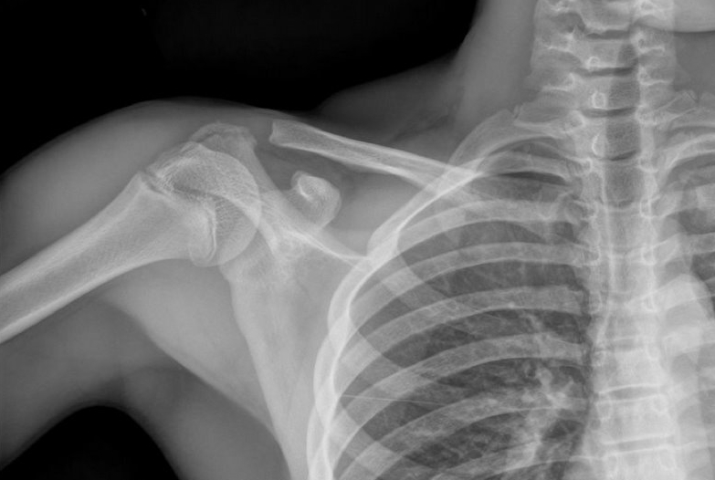
Answer: Non-displaced scapular fracture
Treatment:
Bonus Pearls: #foam4yrdome
This installment of #foam4yrdome will focus on freeemergencytalks.net which is quite possibly the best Critical Care and Emergency Medicine FREE lecture website.
The website was founded and is maintained by Professor Joe Lex (@joelex5); the website hosts hundreds of free talks.
Check out talks from all the major conferences featuring the best speakers in Emergency and Critical care medicine today.
Follow me on Twitter (@criticalcarenow) or Google+ (+criticalcarenow)
Category: Visual Diagnosis
Posted: 7/22/2013 by Haney Mallemat, MD
Click here to contact Haney Mallemat, MD
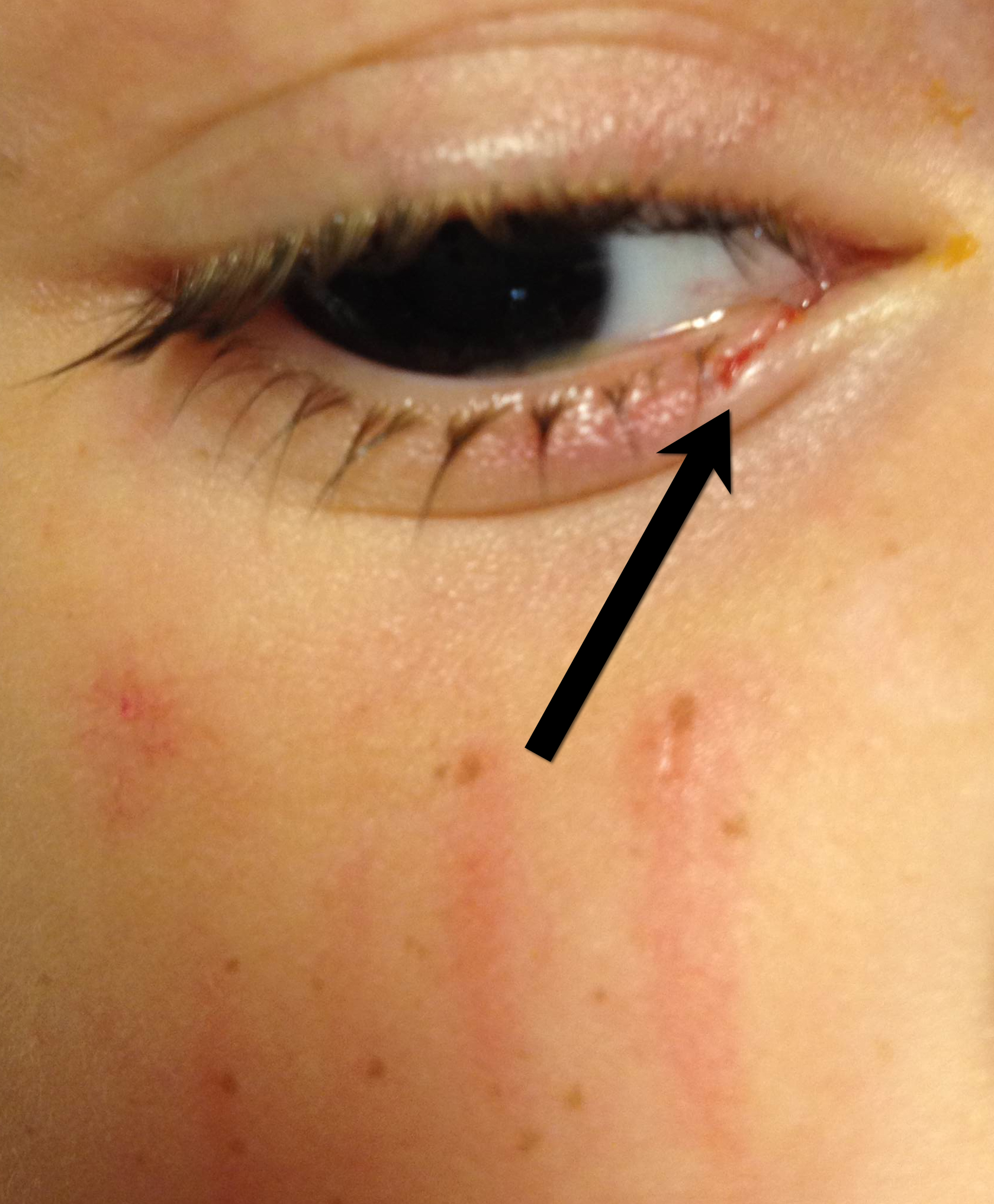
A 3 year-old boy was attacked by a dog and sustained the injury below. Name one injury that should be strongly considered (Hint: there are several)
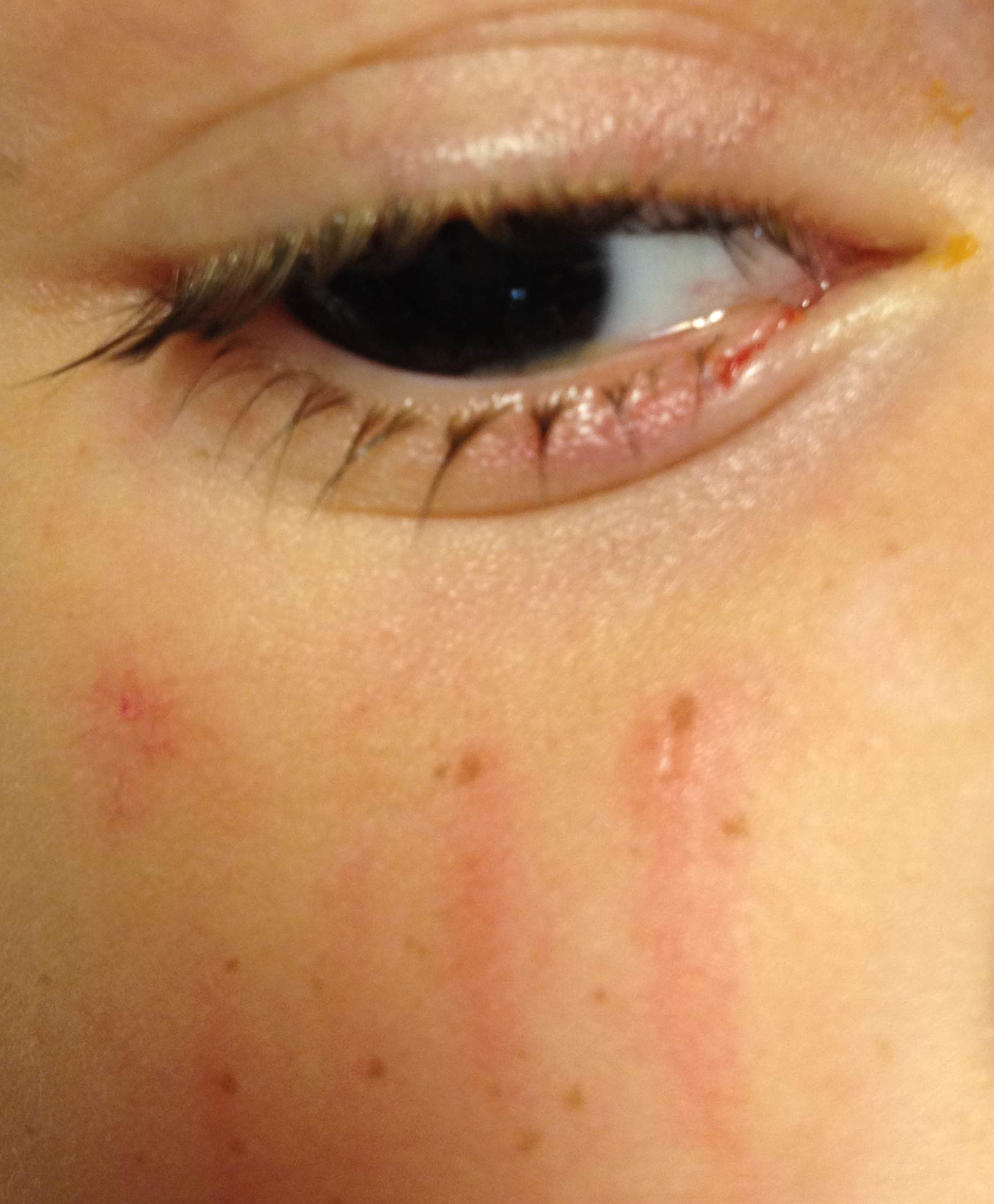
Important injuries to consider (image below):
This patient had only a corneal abrasion on fluorescein exam.
Bonus Pearl: #Foam4yrDome
Follow me on Twitter (@criticalcarenow) or Google+ (+criticalcarenow)
Category: Critical Care
Posted: 7/16/2013 by Haney Mallemat, MD
Click here to contact Haney Mallemat, MD
COPD treatment guidelines (e.g., GOLD) recommend 10-14 days of steroid therapy following a COPD exacerbation to prevent recurrences; the supporting data is weak.
A recent noninferiority trial (here) compared patients with a severe COPD exacerbation who received either a 5-day course (n=156) or 14-day course (n=155) of prednisone 40mg.
The results were:
What you need to know:
Bottom-line: 5 days of prednisone may be as effective as 14-days for COPD exacerbations.
Follow me on Twitter (@criticalcarenow) or Google+ (+criticalcarenow)
Category: Visual Diagnosis
Posted: 7/15/2013 by Haney Mallemat, MD
Click here to contact Haney Mallemat, MD
46 year-old female presents with a headache. The following is seen on visual inspection of the eye. What's the diagnosis?
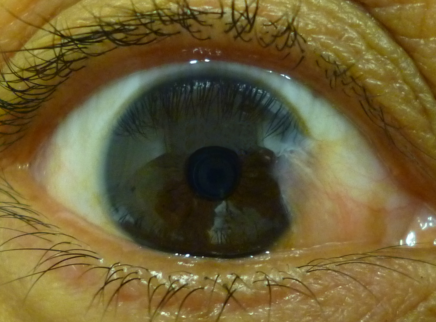
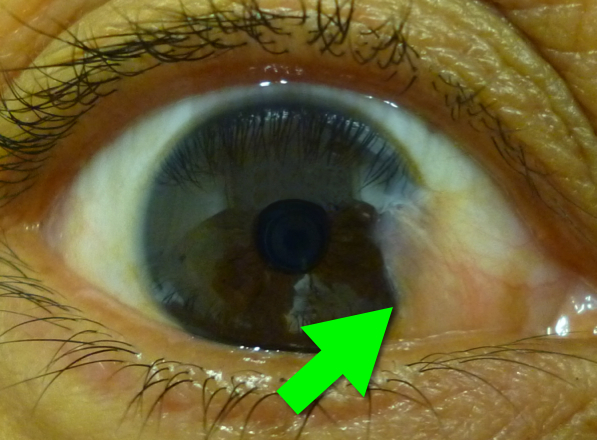
Pterygium
Six-word Summary: Cornea, benign, UV, supportive, surgery, and sunglasses
Bonus Pearl
As a new academic year begins, I will be sharing some amazing free educational online resources/links. These free materials are known as Free Open Access Meducation (or FOAMed) and for those familiar with FOAMed this is an emerging educational revolution. If you don't know what FOAMed is, read about it here and then read this. Updates will happen every Monday and will be known as #FOAM4yourDome
Follow me on Twitter (@criticalcarenow) or Google+ (+criticalcarenow)
Category: Visual Diagnosis
Posted: 7/8/2013 by Haney Mallemat, MD
Click here to contact Haney Mallemat, MD
3 year-old male develops rash 5 days after starting amoxicillin for acute otitis media. What's the diagnosis?

Erythema Multiforme
Erythema multiforme (EM) is a pruritic, erythematous, and blanchable maculopapular rash; it is serpiginous or targetoid in shape, with central clearing or pallor.
EM is generally symmetric, appearing on hands, feet, groin, and extensor aspects of legs and forearms.
It is classically associated with upper respiratory infections, medications, connective tissue diseases, and malignancies.
Treatment includes:
Habif, et al, Skin Disease, 3rd Ed. 2011
Follow me on Twitter (@criticalcarenow) or Google+ (+criticalcarenow)
Category: Critical Care
Posted: 7/2/2013 by Haney Mallemat, MD
Click here to contact Haney Mallemat, MD
Hydroxyethyl starch (HES) is a colloid used for volume resuscitation in critically-ill patients.
Previous studies (click here) have compared crystalloids to HES during fluid resuscitation and have demonstrated that HES has an increased cost with more adverse effects. Adverse effects may include:
In the United States, the Federal Drug Administration published a warning on June 24th 2013 with respect to the use of HES in critically ill adult patients. Specifically, it warned about the use of HES in patients,
If a decision to use HES is made, the FDA warning advises to:
Bottom line: With an increased cost and evidence of harm compared to crystalloids, it appears the indications for use of HES are rapidly declining.
http://www.fda.gov/BiologicsBloodVaccines/SafetyAvailability/ucm358271.htm
Perner A., et al. Hydroxyethyl Starch 130/0.4 versus Ringer's Acetate in Severe Sepsis. NEJM. 2012 Jun 27.
MyBurgh, J. Hydroxyethyl Starch or Saline for Fluid Resuscitation in Intensive Care. N Engl J Med. 2012 Oct 17.
Category: Visual Diagnosis
Posted: 7/1/2013 by Haney Mallemat, MD
Click here to contact Haney Mallemat, MD
65 year-old male presents with nausea and diffuse abdominal pain, 3 days after knee replacement surgery. What's the diagnosis?
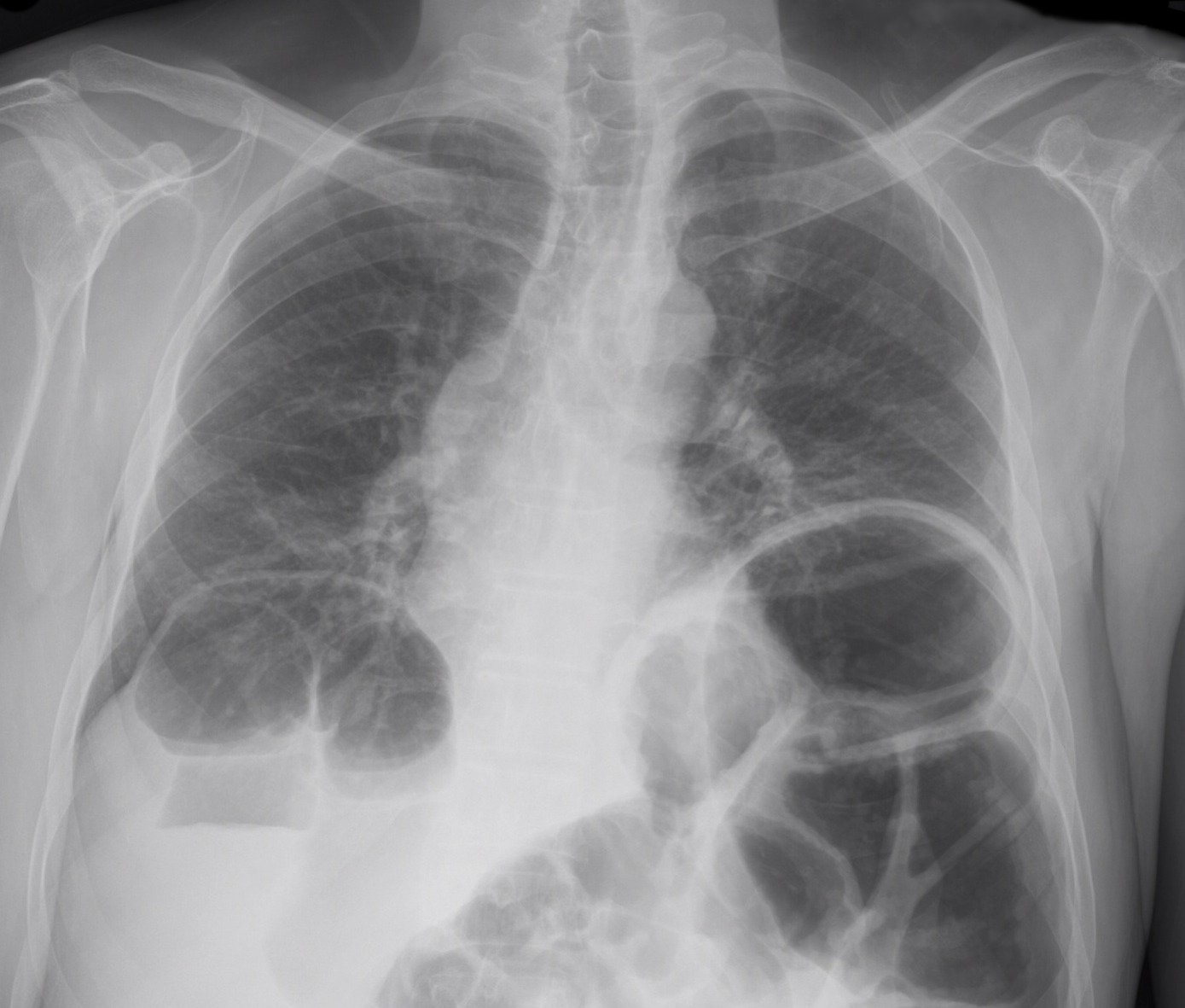
Adynamic ileus
Risk factors include
Imaging reveals distention of both large and small bowel without a transition zone, which differs from a small or large bowel obstruction. Such cases can be difficult to differentiate clinically from one another, so physicians often rely on imaging, specifically CT scanning to define a discrete obstruction versus an ileus.
References
1. Hayden and Sprouse, Bowel Obstruction and Hernia, Med Clin N Amer 29 (2011) 319-345
2. American College of Radiology, Suspected Small Bowel Obstruction, ACR Appropriateness Guidelines, rev. 2010
Follow me on Twitter (@criticalcarenow) or Google+ (+criticalcarenow)
Category: Visual Diagnosis
Posted: 6/23/2013 by Haney Mallemat, MD
(Updated: 6/24/2013)
Click here to contact Haney Mallemat, MD
Name three differential diagnoses based on the CXR below.
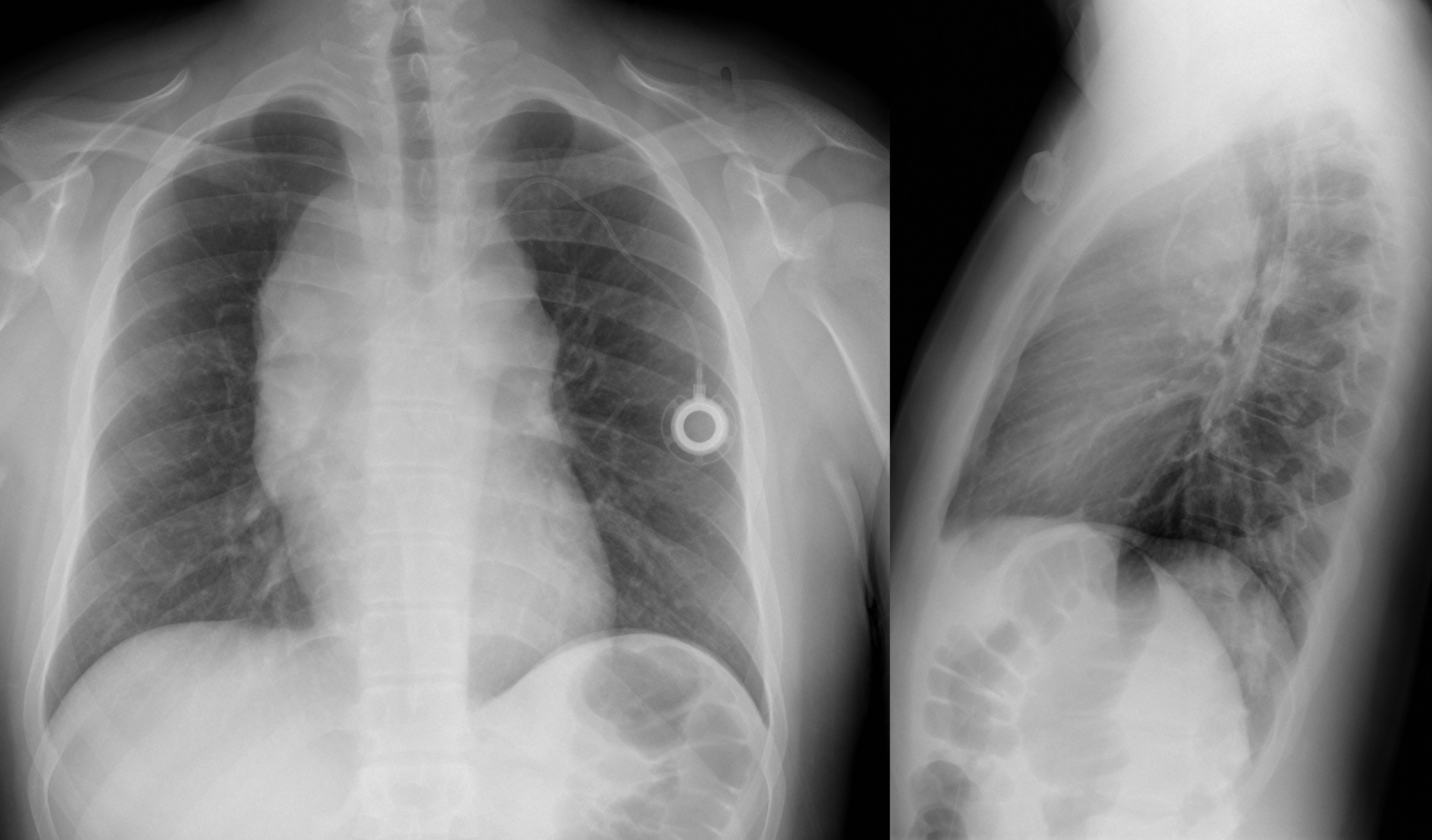
The diagnosis in this case is Non-Hodgkin lymphoma, but read below for more differentials.
Mediastinal Masses
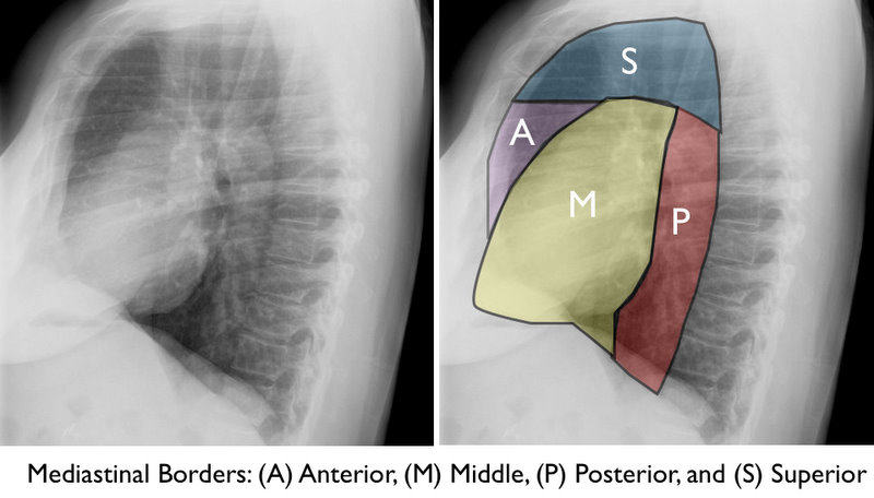
The mediastinum is subdivided into the regions shown below. Here are some differential diagnoses based on region.
Anterior / Superior Mediastinum
Middle Mediastinum
Posterior Mediastinum
Congratulations to all the graduating residents, especially our own at the University of Maryland. I wish you all the best as you start your phenomenal careers!
Follow me on Twitter (@criticalcarenow) or Google+ (+criticalcarenow)
Category: Critical Care
Posted: 6/18/2013 by Haney Mallemat, MD
Click here to contact Haney Mallemat, MD
Keep Immune Thrombocytopenic Purpura (ITP) in your differential for patients with thrombocytopenia and evidence of bleeding. Although ITP has classically been described in children, it can occur in adults; especially between 3rd- 4th decade.
Thrombocytopenia leads to the extravasation of blood from capillaries, leading to skin bruising, mucus membrane petechial bleeding, and intracranial hemorrhage.
ITP occurs from production of auto-antibodies which bind to circulating platelets. This leads to irreversible uptake by macrophages in the spleen. Causes of antibody production include:
Suspect ITP in patients with isolated thrombocytopenia on a CBC without other blood-line abnormalities. Abnormality in other blood-line warrants consideration of another diagnosis (e.g., leukemia).
ITP cannot be cured; treatments include:
