UMEM Educational Pearls - By Michael Bond

Category: Pharmacology & Therapeutics

Title: JNC 8 Recommendations for Hypertension

Keywords: Hypertension, treatment (PubMed Search)

Posted: 12/21/2013 by Michael Bond, MD (Updated: 3/28/2024)
Click here to contact Michael Bond, MD

JNC8 (the Eigth Joint National Commission) released their recommendations for blood pressure management this week. The full article as published in JAMA can be found at http://jama.jamanetwork.com/article.aspx?articleid=1791497

Highlights from this report are

  • Older adults do not need to be placed on antihypertensive medications unless their SBP > 150 or DBP > 90. 
  • Younger patients should still be started if their SBP > 140 or DBP > 90.
  • Firstline drug treatment recommendations are:
    • Non-black patients: start with thiazide diuretics, calcium channel blockers, angiotension converting enzyme (ACE) inhibitors, or angiotension-receptor blockers (ARBs).
    • For black patients start with thiazide diuretics or calcium channel blockers.
    • Patients with chronic kidney disease should be on an ACE or ARB.


General Pearl:  Remember to be cautious in acutely lowering the blood pressure in asymptomatic patients.  Acute lowerings can cause watershed ischemia leading to strokes.


Category: Orthopedics

Title: Posterior Shoulder Dislocation

Keywords: Posterior, Dislocation, Shoulder (PubMed Search)

Posted: 11/30/2013 by Michael Bond, MD (Updated: 3/28/2024)
Click here to contact Michael Bond, MD

Posterior Shoulder Dislocations

  • A rare type of shoulder dislocation
  • Accounts for 2-4% of all shoulder dislocations
  • Classic mechanism of injury is a seizure or electrocution
  • Reported to occur bilaterally in 15% of cases
  • Often missed on the initial visit.
  • Patient will complain of pain with movement of the shoulder and the arm is held in internal rotation.
  • Can be missed on the AP, lateral and Y-views of the shoulder.
  • Axillary or modified Axillary views are the best view to visualize a posterior shoulder dislocation. Shown below:

Axillary View of Shoulder

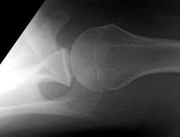
(A posterior shoulder dislocation will show the humeral head displayed superiorly in the image away from the clavicle which is the inferior most bone)

Some things to look for on the AP view that will suggest a posterior shoulder dislocation:

  • Lightbulb sign – The head of the humerus in the same axis as the shaft producing a lightbulb shape
  • The ‘rim sign’ – Widening of the glenohumeral space
  • The vacant glenoid sign – the anterior glenoid fossa appears empty

Life in the Fast Lane as a great discussion of posterior shoulder dislocations at http://lifeinthefastlane.com/posterior-shoulder-dislocation/

 Best way to make the diagnosis --- suspect it and get an axillary view.


Category: Orthopedics

Title: Compartment Syndrome

Keywords: Compartment Syndrome (PubMed Search)

Posted: 11/16/2013 by Michael Bond, MD (Updated: 3/28/2024)
Click here to contact Michael Bond, MD

Compartment Syndrome

Compartment syndrome is classically described as having the 6 Ps:

  • Pain out of proportion to what is expected
  • Pulselessness [Late finding that you hope to never see]
  • Paresthesia
  • Paralysis
  • Pallor
  • Pressure


The diagnosis of compartment syndrome can be difficult but ultimately it comes down to measuring the pressures in the area of concern.  Various recommendations of the allowed pressure can be found, but in general a fasciotomy is not needed if the compartment pressure is 30 mmHg less then the diastolic pressure (The Delta 30).  So if the patients diastolic pressure is 70, a fasciotomy is not need if the compartment pressure is less then 40.  

Finally, if you are suspecting compartment pressure do NOT elevate the limb.  Leave it in a dependent position to help improve blood flow into the limb.


As many pregnant and previously pregnant women will tell you, the term morning sickness is really a misnomer. Nausea and vomiting can really occur at any time of day (and often does). The mechanisms through which this happens is really unknown. Limited research suggests that the placenta may be responsible for the symptoms given that patients with hydatidiform molar pregnancies (no fetus) have some of the most severe cases of nausea and vomiting. 
 

Treatment:

  • Vitamin B6 has been shown in a number of randomized placebo-controlled trials to be very effective in treatment of the 1st trimester nausea and vomiting and is a supported recommendation by the American College of Obstetrics and Gynecology (ACOG).
    • For refractory symptoms, efficacy is increased with supplementation of the sleep aide Doxylamine (found in Unisom pills over the counter)
    • Dosing of Vitamin B6 is 10 to 25mg every 8 hours daily
    • Dosing for Doxylamine is 12.5mg as needed in the morning, 12.5mg as needed in the afternoon, and 25mg as needed at bedtime.
  • Ginger (ginger ale and ginger supplements) taken at 250mg daily doses has been recommended as 1st line treatment by ACOG.

Consider these therapies the next time you see a pregnant with persistent nausea and vomiting in her 1st


--Yemi

Show References


Category: Orthopedics

Title: Charcot Joints

Keywords: Charcot Joints (PubMed Search)

Posted: 8/17/2013 by Michael Bond, MD
Click here to contact Michael Bond, MD

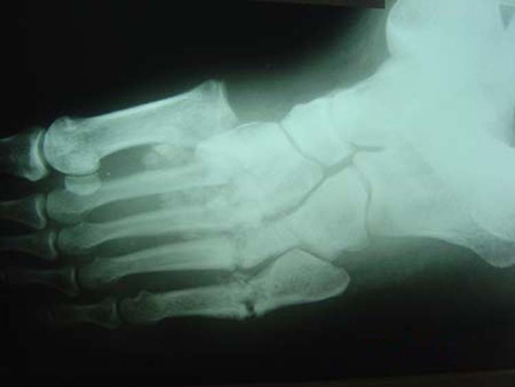
Charcot Joint - Neuropathic arthropathy

A Charcot Joint is a progressive degeneration of a weight bearing joint that is normally seen in patients that have decreased peripheral sensation and proprioception.

Conditions associated with Charcot Joints are:
    •    Alcohol neuropathy
    •    Cerebral palsy
    •    Diabetes mellitus
    •    Spinal Cord Injury
    •    Strokes
    •    Syphilis (tabes dorsalis)

The foot is most commonly affected and radiographs can also show bony destruction, bone resorption, and gross deformity. The onset of pain and deformity is typically insidious.  Charcot joints are often associated with ulcerations, secondary osteomyelitis, and can lead to amputations.

Charcot Joint

It is important to recognize the presence of a Charcot Joint so that the patient can be referred to Orthopaedics and treated (often with cast immobilization) to prevent further destruction of the joint.


Category: Endocrine

Title: Treatment of Severe Hypothyroidism

Keywords: Hypothyroidism, treatment (PubMed Search)

Posted: 7/20/2013 by Michael Bond, MD (Updated: 3/28/2024)
Click here to contact Michael Bond, MD

Treatment of Severe Hypothyroidism

We do not see patient's with severe hypothyroidism often, but it is important that they be treated aggressively. Some treatment pearls are

  • Rule out aggravating cause (i.e.: infection [UTI, pneumonia], myocardial infarction)
  • Start IV levothyroxine dosing
    • Initial dose 400-500 mcg. This is a large dose but it only saturates the thyroid receptors and will not cause a rebound hyperthyroidism state.
    • Daily dose 100 mcg/day
  • Consider starting Dexamethasone/hydrocortisone
    • Patients may also have adrenal insufficiency from primary pituitary failure or may have secondary adrenal suppression due to the severe hypothyroidism.  If dexamethasone/hydrocortisone is not provided they may develop severe adrenal insufficiency once you kick start their metabolism.


Category: Orthopedics

Title: Sternal Fractures

Posted: 6/29/2013 by Michael Bond, MD (Updated: 3/28/2024)
Click here to contact Michael Bond, MD

Sternal fractures

  • Initially thought to be associated with high mortality due to associated injuries though newer studies show the mortality rate is about 1%.
  • Can be associated with
    • Rib fractures
    • Mediastinal injury
    • Cardiac Contusion
    • Pneumothorax
    • Aortic dissection
    • Pulmonary Contusion
  • The diagnosis can be made with plain radiographs, but a fracture can be missed on a regular PA and Lateral Chest Xray.  Ask for dedicated sternal views to better define the fracture
    • CT Chest is only needed if you are concerned about associated injuries
  • Obtain an ECG on arrival and at 6 hours to ensure there are no signs of a myocardial contusion
    • ST segment changes, arrhthymias
  • Treatment is supportive. Provide adequate pain control and treat associated injuries

Show References


Category: Orthopedics

Title: Trapezium Fractures

Keywords: Trapezium, Fracture (PubMed Search)

Posted: 6/15/2013 by Michael Bond, MD
Click here to contact Michael Bond, MD

Trapezium Fractures

  • The trapezial body is often fractured due to an axial load through the thumb
  • The trapezial ridge is often fractured during a fall on an outstretched hand
  • Accounts for 1% to 5% of all carpal fractures.

Suspect the Diagnosis when you note

  • Tenderness  over trapezium 
  • Often have increased pain with axial loading of thumb.
  • Point tenderness at the volar base of the thumb just distal to the scaphoid, at the base of the first metacarpal.
  • Since the trapezium is obscured by superimposed bones in PA and lateral views, fractures are most easily identified on the oblique radiographs

If you are suspected the diagnosis oblique radiographs or a CT scan of the wrist will note the fracture the best.

Treatment consists of placing the patient in a thumb spica splint.


Category: Orthopedics

Title: Fabella Syndrome

Keywords: Fabella (PubMed Search)

Posted: 5/18/2013 by Michael Bond, MD (Updated: 3/28/2024)
Click here to contact Michael Bond, MD

Fabella Syndrome

The fabella is a sesamoid bone that is embedded in the tendon of the gastrocnemius muscle where the fibers of the popliteus, arcuate complex and the fibular-fabellar ligament attach.

Fabella syndrome is a painful condition of the posterolateral knee that is exacerbated when the knee is extended.  The pain can be exacerbated by palpation of the fabella and if it is compressed against the condyles. The condition is most common in adolescence, but occurs in adults too.

Consider this condition in patients with posterolateral knee pain, which can also be due to tears of the  posterior horn of the lateral meniscus, and tendonitis of the lateral head of the gastrocnemius.


Category: Orthopedics

Title: Knee Injuries are Radiographs Needed

Keywords: Ottawa, Knee, Pittsburgh (PubMed Search)

Posted: 3/30/2013 by Michael Bond, MD
Click here to contact Michael Bond, MD

Knee Pain Injuries are Radiographs needed?


Many people know that the folks in Ottawa have come up with a rule to determine whether radiographs are needed in patients complaining of knee pain.  The Ottawa Knee rules that that radiographs are only required for knee injuries with any of the following:
    •    Age 55 years or older
    •    isolated tenderness of patella
    •    tenderness at head of fibula
    •    inability to flex to 90'
    •    inability to bear weight both immediately and in the emergency department (4 steps)

Well another group in Pittsburgh have their own set of rules that were recently shown to be more specific with equal sensitivity.  The Pittsburgh decision rules state that radiographs are only needed if

  • There is a history of fall or blunt trauma AND  ( Patient is < 12 or > 50 years old OR Patient is unable to walk for weight bearing steps in the ED. )

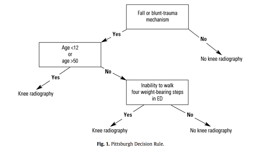
So consider using the Pittsburgh or Ottawa Knee rules the next time you have a patient with knee pain to determine if those radiographs are really needed.

The full article can be found at http://www.ajemjournal.com/article/S0735-6757%2812%2900566-9/abstract

Show References


Category: Orthopedics

Title: Board Review - Scapular Fractures

Keywords: scapular, fracture (PubMed Search)

Posted: 3/16/2013 by Michael Bond, MD (Updated: 3/28/2024)
Click here to contact Michael Bond, MD

Scapular fractures

  • Usually the result of a significant force, because of this associated injuries are frequent and sometimes life- or limb-threatening.
  • Some of the associated injuries are:
  • Rib fractures
  • Ipsilateral lung injuries
  • Pneumothorax
  • Hemothorax
  • Pulmonary contusion
  • Injuries to the shoulder girdle complex
  • Clavicle fractures
  • Shoulder dislocations with associated rotator cuff tears
  • Neurovascular injuries (rare)
  • Brachial plexus injuries
  • Axillary artery or nerve injuries
  • Subclavian artery injury
  • Suprascapular nerve injury
  • Vertebral compression fractures

Show References


Category: Orthopedics

Title: Boxer's (Metacarpal Neck) Fractures

Keywords: metacarpal, neck, fracture (PubMed Search)

Posted: 12/29/2012 by Michael Bond, MD
Click here to contact Michael Bond, MD

Metacarpal Neck Fractures (i.e.: Boxer’s Fracture if 5th Metacarpal)

Depending on the MCP joint involved a certain amount of angulation is permissible before it adversely affects normal function.

  • 2nd and 3rd Metacarpal fractures < 10۫ angulation ideally these should be perfectly aligned.
  • 4th Metacarpal fracture <20۫ angulation allowed
  • 5th Metacarpal fracture <30۫ angulation.
    • Studies have shown that even 30۫ angulation will decrease normal function by 20%.  
    • Normal excursion of the 5th MCP is 15۫ to 25۫.
  • No amount of rotation deformity should be allowed.

Wishing everybody a Happy and Healthy New Year.


Category: ENT

Title: Epistaxis Control

Keywords: epistaxis (PubMed Search)

Posted: 12/15/2012 by Michael Bond, MD
Click here to contact Michael Bond, MD

Epistaxis can be a difficult thing to control in the ED, but there are several techniques you can learn that will make your life easier.

The majority of epistaxis cases are from kiesselbach's plexus therefore you can control it with:

Direct Pressure: Can be held with two fingers pinching the nares, or you can tape 4 tongue blades together and make your own "clothes pin" that can then be used to pinch the nares.

Vasoconstrictor and Anesthesia: A 1:1 mixture of topical lidocaine 4% and oxymetazoline can often be mixed together in the same oxymetazoline spray container enabling you to just spray it into the nares. This will often slow or stop the bleeding and provides anesthesia in case you need to cauterize the bleeding site.  Some IV/IM narcotic pain medication will also help increase patient cooperation.

Visualize the bleeding site: Use a HEAD LAMP with an appropriate sized nasal speculum. You may look like Marcus Welby, MD but nothing works as well to see into the nose.

Cauterization It is best to cauterize circumferential around the bleeding site prior to directly cauterizing the actual site. Be careful with electrical cautery so has not to perforate the septum.

Nasal Packing: Instead of using surgilube to lubricate the packing; use Muprion, Bactroban or Bacitracin ointment to lubricate the packing. This will reduce the chance of Toxic Shock Syndrome.


Category: Orthopedics

Title: Tarsal Tunnel Syndrome

Keywords: tarsal tunnel syndrome (PubMed Search)

Posted: 11/17/2012 by Michael Bond, MD (Updated: 3/28/2024)
Click here to contact Michael Bond, MD

Tarsal Tunnel Syndrome (TTS)

Prior pearls have addressed Carpal Tunnel Syndrome and Cubital Tunnel Syndrome, which affect the median and ulnar nerves, respectively.  Tarsal tunnel syndrome, is a similar compression neuropathy of the tibial nerve as it transverses through the tarsal tunnel of the foot.

The tarsal tunnel is located behind the medial malleolus, and is where the posterior tibial artery, tibial nerve and several tendons transverse.  Patients will present complaining of numbness of the foot radiating into  Digits 1-4, pain, burning , and tingling of the base of the foot and heel.  TTS has many causes and is more common in athletes.

Consider the diagnosis in patients with foot pain and numbness.  If interested in more information about TTS please consider reading this eMedicine article, http://emedicine.medscape.com/article/1236852-overview


Fight Bites

  • Any abrasion or laceration over the knuckles should be presumed to be a fight bite.  Patients will often lie about the circumstances of the laceration.
  • Radiographs should be obtained on all of these patients to exclude
    • Retained foreign bodies (e.g., tooth fragments)
    • Fracture of the metacarpal head
  • Place patient on amoxicillin/clavulanic acid or clindamycin to cover mouth flora
  • Irrigate wound well, and explore through the fingers full range of motion to exclude joint or tendon injury.
  • Refrain from suturing the wound, as this will increase the risk of infection.
  • Splint in position of function and have them follow up in 2 days.


Lactate levels help to confirm septic arthritis but what about bacterial meningitis.  As reported in the daily electronic ACEP newsletter a small study of 45 patients showed that all patients with a confirmed diagnosis of bacterial meningitis had a CSF lactate level > 3.5 mmol/L.  Therefore, it might be true that viral meningitis will only have  CSF lactate levels < 3.5 mmol/L. 

With only 45 patients, this finding is clearly not ready for Prime Time but consider adding it to your next CSF study so more data can be collected on the utility of this test.

The story as seen in ACEP eNews on September 14th, 2012 is:

CSF Levels Of Lactate May Be A Marker Of Viral Versus Bacterial Meningitis.

MedPage Today (9/14, Gever) reports, "Cerebrospinal fluid (CSF) levels of lactate were a perfect marker of viral versus bacterial meningitis in a small study, a researcher reported" at the Interscience Conference on Antimicrobial Agents and Chemotherapy. Researchers found that, "among 45 adults in whom the etiology of meningitis was microbiologically confirmed, all those with CSF lactate levels above 3.5 mmol/L had the bacterial form, whereas every patient with lower levels had viral meningitis."


Category: Orthopedics

Title: Synovial Fluid Analysis

Keywords: lactate, synovial fluid, (PubMed Search)

Posted: 8/18/2012 by Michael Bond, MD (Updated: 3/28/2024)
Click here to contact Michael Bond, MD

The Analysis of Synovial Fluid Analysis

When trying to diagnosis a septic joint, it is common to order the following labs on the synovial fluid:

  • Synovial WBC count
  • Glucose
  • Protein
  • Gram Stain
  • Culture

Unfortunately, there is no value of glucose or protein that has enough sensitivity and specificity to make the tests diagnostically helpful.  Gram stains are only postive in culture positive septic joints in approximately 50% of the cases.  Cultures take too long to be helpful in the ED.  The synovial WBC count can be helpful if very high, but a low value does not ensure that the patient does not have a septic joint.

The one test that has been shown to have a Positive Likelihood ratio of Infinity is a synovial lactate level >10.  A synovial lactate should be sent on all synovial fluid as a level of 10 and greater makes the diagnosis of septic arthritis, regardless of the gram stain or synovial WBC level.


Show References


Category: Orthopedics

Title: Humerus Fractures

Keywords: Humerus Fractures (PubMed Search)

Posted: 7/21/2012 by Michael Bond, MD (Updated: 8/28/2014)
Click here to contact Michael Bond, MD

Humerus Fractures, Proximal

  • Proximal fractures of the humerus only account for about 5% of all fractures but account for 45% of all humeral fractures.
  • We should be familar with the Neer Classication System for Humeral fractures, which can also be seen at http://health-7.com/Handbook%20of%20Fractures/15%20-%20Proximal%20Humerus%20Fractures
     
  • The classification system classifies fractures based on
    • 1-part
    • 2-part
    • 3-part
    • 4-part
  • The bony segments that make up the parts are
    • Greater Tuberosity (GT)
    • Lesser Tuberosity (LT)
    • Humeral Head
    • Humeral Surgical Neck (SN)
  • A part is defined as displaced if >1 cm of fracture displacement or >45 degrees of angulation.
  • The greater the number of parts the more likely the patient will require surgery or have increased complications.
    • 3 and 4 part fractures are often fixed surgical due to the increased risk of vascualr compromise to the humeral head.


Category: Orthopedics

Title: First Metacarpal Fractures

Keywords: Bennett, Rolando, fracture (PubMed Search)

Posted: 6/30/2012 by Michael Bond, MD (Updated: 3/28/2024)
Click here to contact Michael Bond, MD

First Metacarpal Fractures:

There are two types of fractures that commonly occur at the base of the 1st metacarpal.  They are:

Bennett Fracture:  This is an intraarticular fracture at the base of the 1st metacarpal that always involves some degree of subluxation or dislocation of the 1st carpometacarpal joint. 

Bennett Fracture

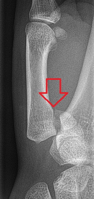
Image from Wikipedia Commons

  • This is the most common fracture of the thumb. 
  • This fracture is typically caused by axial loading the thumb while the metacarpal is partially flexed.  A common mechanism is when a person punches a wall or hard object with their thumb take the brunt of the force.
  • Most of these fractures will require surgical repair but can be treated in the ED with a Thumb Spica Splint.

Rolando Fracture:  This is a communited intraarticular fracture at the base of the first metacarpal that typically has a T or Y shaped configuration with 3 fragments.

Rolando Fx

Image courtesy of WikiPedia Commons

  • Less common then Bennett Fractures
  • Associated with a worse prognosis
  • Requires surgical repair but can be splinted in the ED with a thumb spica splint.


Category: Misc

Title: Contrast Allergy

Keywords: contrast media, iodine, shellfish (PubMed Search)

Posted: 6/16/2012 by Michael Bond, MD
Click here to contact Michael Bond, MD

Contrast Allergy:

Many patients will report that they have a allergy to iodinated contrast by saying that they are allergic to iodine

Iodine, itself, is not an allergen and is a required element for thyroid homrone production.  Plus could you imagine the hordes of people that would be having allergic reactions everyday when they add salt to their french fries.  Our EDs would be completely swamped.

A recent meta-analysis by Drs. Schabelman and Witting also showed the following:

  • The risk of a reaction to contrast ranges from 0.2% to 17% depending on the type used, and the severity of the reaction considered.
  • The risk of a reaction in patients with a seafood allergy is similar to that in patients with other food allergies or asthma.  Seafood is not unique to contrast media.
  • A history of prior reaction to contrast increases the risk of mild reactions to as high as 7-17% but has not been shown to increase the rate of severe reactions.
  • The risk of death due to contrast is estimated to be 0.0006 - 0.006%.

As we enter Crab eating season in Maryland, lets stop giving shellfish a bad name. A patent with any allergy is at increased risk, but shellfish is no higher a risk than those allergic to Strawberries.

Show References