A 50 years old male with a history of CHF, presenting to the ED with progressively worsening shortness of breath. POCUS was performed. The picture shows the left lower part of the chest. What is the diagnosis?
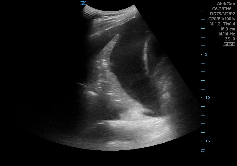
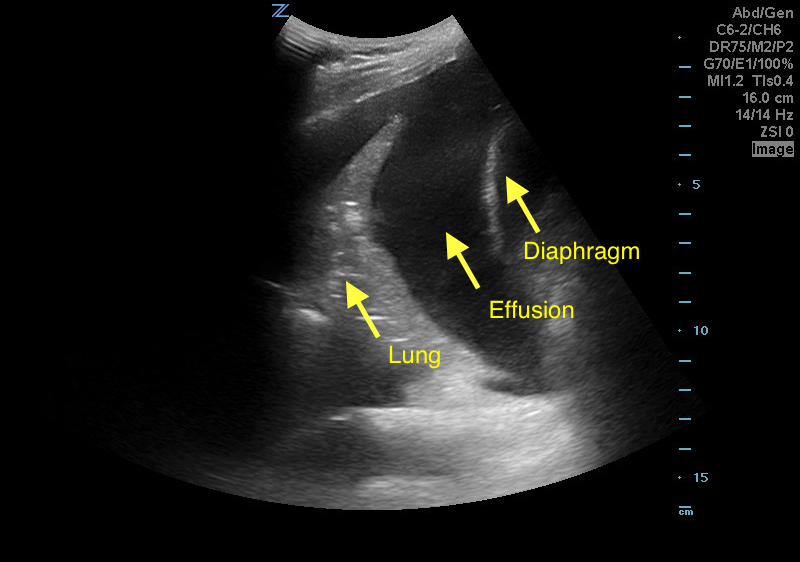
Answer: Pleural effusion
Eibenberger, K. L., Dock, W. I., Ammann, M. E., Dorffner, R., Hörmann, M. F., & Grabenwöger, F. (1994). Quantification of pleural effusions: sonography versus radiography. Radiology, 191(3), 681-684.
Atkinson, P., Milne, J., Loubani, O., & Verheul, G. (2012). The V-line: a sonographic aid for the confirmation of pleural fluid. Critical ultrasound journal, 4(1), 19.