Category: Visual Diagnosis
Posted: 5/5/2014 by Haney Mallemat, MD
Click here to contact Haney Mallemat, MD
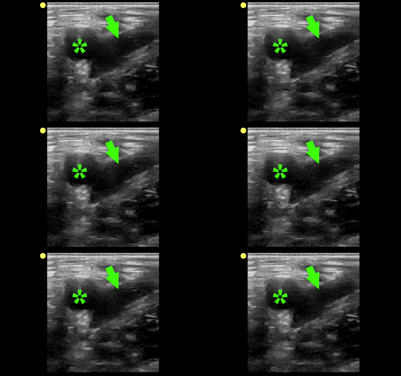
The clip below demonstrates normal right femoral anatomy. The structure with the asterisk is the right common femoral vein and the arrow is pointing to a branch of the right femoral vein. What is the name of the branch and what is its importance during lower extremity ultrasound?

Answer: Greater Saphenous Vein; it is one of the two regions that should be compressed when evaluating for a lower extremity DVT in the Emergency Department (the other is at the trifurcation of the popliteal vein).
Here is a podcast from the Ultrasound Podcast describing the entire bedisde DVT exam http://www.ultrasoundpodcast.com/2011/08/dvt/
Follow me on Twitter (@criticalcarenow) or Google+ (+criticalcarenow)a
