Category: Orthopedics
Keywords: Subungual Hematomas (PubMed Search)
Posted: 10/16/2010 by Michael Bond, MD
(Updated: 2/8/2026)
Click here to contact Michael Bond, MD
Subungual Hematomas:
Category: Orthopedics
Keywords: joint, documentation, physical examination (PubMed Search)
Posted: 10/9/2010 by Brian Corwell, MD
(Updated: 2/8/2026)
Click here to contact Brian Corwell, MD
Previous pearls have described tips for smart and safe documentation of typical ED complaints such as chest pain. Properly assessing and documenting orthopedic complaints is likewise very important. No evaluation or chart is complete if it does not include include the following 7 components:
The joint above
The joint below
Motor
Sensory
Vascular
Skin
Compartments
The joint above/below is important in cases of shoulder and hip pain actually being radicular pain (from the neck and back respectively). Also, hip pain from trauma may be due to a femur fracture for example.
For motor and sensory evaluation, test the most distal isolated innervation of a particular nerve (L5 - great toe dorsiflexion for example).
Note distal pulses and check ABIs for injuries with potential subtle vascular findings.
Note intact skin especially in cases where the joint will be covered by a splint.
Note "soft" compartments especially in cases of forearm and lower leg fractures.
Category: Orthopedics
Keywords: Patellofemoral syndrome (PubMed Search)
Posted: 10/2/2010 by Michael Bond, MD
Click here to contact Michael Bond, MD
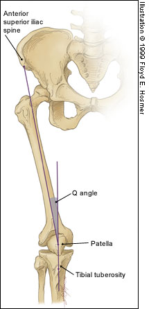
Patellofemoral Syndrome (Chondromalacia Patella)

Juhn MS et al. Patellofemoral pain syndrome: a review and guidelines for treatment. Am Fam Physician. (1999)
Category: Orthopedics
Keywords: Thumb, Gamekeeper's thumb, Skier's thumb (PubMed Search)
Posted: 9/25/2010 by Brian Corwell, MD
(Updated: 9/28/2010)
Click here to contact Brian Corwell, MD
Injury was originally described as an occupational hazard in Scottish gamekeepers (from breaking the necks of rabbits against the ground). Today, skiing is now the most common cause and injury is now the second most common orthopedic injury in skiers (MCL injury #1).
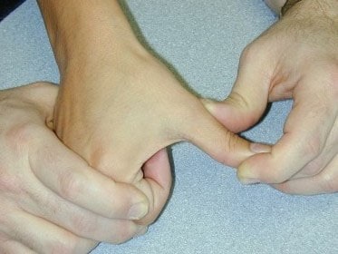
Injury to the ulnar collateral ligament (UCL) results from a sudden forced abduction (radial deviation) stress at the MCP joint of the thumb, commonly due to a fall against a ski pole or the ground.

http://blog.fitter1.com/wp-content/uploads/2010/04/b_14_1_2a.jpg
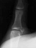
The most frequent site of rupture is the insertion into the proximal phalanx. The UCL may even avulse a small portion of the proximal phalanx at its insertion site.

http://img.medscape.com/pi/emed/ckb/sports_medicine/84611-97564-98460-1652013.jpg
Consider imaging before stress testing (to avoid further displacing a fracture)

http://img.medscape.com/pi/emed/ckb/sports_medicine/84611-97564-98460-1652060.jpg
Stabilize in a thumb spica splint and refer to hand surgery.
Calling this entity a “simple sprain” may result in chronic disability (chronic pain, instability, loss of pinch strength)
http://emedicine.medscape.com/
Category: Orthopedics
Keywords: Pain, Geriatrics (PubMed Search)
Posted: 9/18/2010 by Michael Bond, MD
(Updated: 2/8/2026)
Click here to contact Michael Bond, MD
Pain Control in the Elderly
So the take home lesson for this pearl is that the elderly have a lower risk of delirium if their pain is treated appropriately.
Duggleby W, Lander J: Cognitive status and postoperative pain: older adults. J Pain Symptom Manage 1994; 9: 19-27.
Lynch EP, Lazor MA, Gellis JE, Orav J, Goldman L, Marcantonio ER: The impact of postoperative pain on the development of postoperative delirium. Anesth Analg 1998; 86: 781-785.
Category: Orthopedics
Keywords: Shoulder, Rotator cuff (PubMed Search)
Posted: 9/11/2010 by Brian Corwell, MD
(Updated: 12/18/2010)
Click here to contact Brian Corwell, MD
Supraspinatus: “Empty can” test. Have the patient abduct the shoulders to 90 degrees in forward flexion with the thumbs pointing downward. The patient attempts to lift the arms against the examiner’s resistance.
http://bjsportmed.com/content/42/8/628/F2.large.jpg
Infraspinatus and teres minor: These muscles are responsible for external rotation of the shoulder. Have the patient flex both elbows to 90 degrees while the examiner provides resistance against external rotation.
http://www.physio-pedia.com/images/4/4b/Infraspinatus_test.jpg
Subscapularis: “Lift-off” test. The patient rests the dorsum of the hand on the lower back (palm out) and then attempts to move the arm and hand off the back. Patients with tears may be unable to complete test due to pain.
http://www.aafp.org/afp/2008/0215/afp20080215p453-f4.jpg
1) http://bjsportmed.com
2) http://www.aafp.org
Category: Orthopedics
Keywords: Elbow, radiographs (PubMed Search)
Posted: 9/4/2010 by Brian Corwell, MD
Click here to contact Brian Corwell, MD
Radiologic evaluation of the elbow (Part 2)
Helpful clues in the evaluation of elbow trauma:
Category: Orthopedics
Keywords: Adhesive Capsulitis (PubMed Search)
Posted: 8/28/2010 by Michael Bond, MD
Click here to contact Michael Bond, MD
Adhesive Capsulitis -- Frozen Shoulder
Category: Orthopedics
Keywords: Rotator Cuff Tears, Chronic, Acute (PubMed Search)
Posted: 8/21/2010 by Michael Bond, MD
Click here to contact Michael Bond, MD
Rotator Cuff Tears:
Four muscles make up the rotator cuff (SITS) which control internal and external rotation of the shoulder and abduct the shoulder.
Tears can be due to acute injuries (falls, heavy lifting, forceful abduction), though the majority (>90%) of rotator cuff tears are chronic in nature and due to subacromial impingement and decreased blood supply to the tendons.
Most patients can be treated with sling immobilization, NSAIDs and referral to sports medicine or orthopaedic surgeons. Elderly patients should be referred quickly as prolonged immobilization can lead to a frozen shoulder.
Category: Orthopedics
Keywords: Elbow, fat pad, fracture (PubMed Search)
Posted: 8/14/2010 by Brian Corwell, MD
(Updated: 9/18/2010)
Click here to contact Brian Corwell, MD
Helpful clues in the evaluation of elbow trauma
Fat pads: The fat pad sign can be seen with any joint effusion (infection, inflammation) but in the setting of trauma, effusions are indicative of fractures about the elbow (even if no fracture line can be identified).
There are two fat pads within the elbow. Normally, on a true lateral radiograph only the anterior fat pad is seen as a small triangular radiolucent shadow anterior to the distal humeral diaphysis. The posterior fat pad is ordinarily not visualized on a lateral radiograph because it is tucked away within the olecranon fossa.
Normal lateral view: http://nypemergency.org/images/ElbowNormal.jpg
With fractures, the joint becomes distended with blood. The anterior fat pad becomes displaced superiorly and outward from the humerus giving the so called "sail sign." Similarly, the posterior fat pad gets displaced out of the olecranon fossa and becomes visible on the lateral radiograph.
Anterior (sail) and posterior fat signs: http://nypemergency.org/images/Elbowsfatpadarrow.jpg
http://nypemergency.org
Category: Orthopedics
Posted: 8/7/2010 by Michael Bond, MD
(Updated: 2/8/2026)
Click here to contact Michael Bond, MD
Some common injuries and their board review associated complications
Category: Orthopedics
Posted: 7/24/2010 by Brian Corwell, MD
(Updated: 2/8/2026)
Click here to contact Brian Corwell, MD
| History and Physical Examination Red Flags | |
| Historical Red Flags | Physcial Red Flags |
| Age under 18 or over 50 Pain lasting more than 6 weeks History of cancer Fever and chills Night sweats, unexplained weight loss Recent bacterial infection Unremitting pain despite rest and analgesics Night pain Intravenous drug users, immunocompromised Major trauma Minor trauma in the elder | Fever Writhing in pain Bowel or bladder incontinence Saddle anesthesia Decreased or absent anal sphincter tone erianal or perineal sensory loss Severe or progressive neurologic defect Major motor weakness |
Category: Orthopedics
Keywords: Salter Harris (PubMed Search)
Posted: 7/17/2010 by Michael Bond, MD
Click here to contact Michael Bond, MD
The Salter Harris Classification System is used in pediatric epiphyseal fractures. The higher the type of fracture the poorer the prognosis
Some common exam facts about Salter Harris Fractures are:
The Classification system as listed by Type:
A image of the fractures can be found on FP Notebook at http://www.fpnotebook.com/_media/OrthoFractureSalterHarris.jpg
Category: Orthopedics
Keywords: Spondylolysis (PubMed Search)
Posted: 7/10/2010 by Brian Corwell, MD
(Updated: 2/8/2026)
Click here to contact Brian Corwell, MD
http://www.gentili.net/signs/images/400/spinescottyparsdefectdrawing.JPG
The Scotty dog’s head (superior articular facet), nose (transverse process), eye (pedicle), neck (pars interarticularis), and body (lamina) should be easily identified on the oblique radiograph.
Category: Orthopedics
Keywords: Odontoid, fracture (PubMed Search)
Posted: 6/26/2010 by Michael Bond, MD
(Updated: 2/8/2026)
Click here to contact Michael Bond, MD
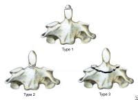
Odontoid Fractures:
There are three types of C2 odontoid fractures:

Category: Orthopedics
Keywords: Calcaneus Fracture, Bohler Angle (PubMed Search)
Posted: 6/13/2010 by Michael Bond, MD
Click here to contact Michael Bond, MD
Calcaneus Fractures:
Calcaneus fractures can easily be missed on plain films and the true extent of the injury might not be appreciated until a CT is done. However, you can increase your change of picking up a calcaneal fracture by evaluating Bohler's Angle.
Lateral radiographs of the foot are needed to evaluate the Bohler angle. This is the angle made by drawing a line from anterior process of the calcaneus to the peak of the posterior articular surface and a second one drawn from the peak of the posterior articular surface to the peak of the posterior tuberosity. (See Picture) The average angle is 25-40°. Angles less than 25' are strongly suggestive of a fracture and the patient should probably get a CT of their foot if there is clinical suspicion.

Photo and additional discussion can be found at http://www.blackburnfeet.org.uk/hyperbook/trauma/calcanealFx/imaging.htm and on eMedicine -- Calcaneus Fractures by Scott Nicklebur, MD http://emedicine.medscape.com/article/1232246-overview
Category: Orthopedics
Keywords: Wound Care, Antiseptics (PubMed Search)
Posted: 6/5/2010 by Michael Bond, MD
Click here to contact Michael Bond, MD
Wound Care:
Patients and many providers want to irrigate or wash a wound with an antiseptic solution in order to decrease the risk of infection. Most studies have shown that irrigation whether with tap water or sterile water is effective enough in reducing bacterial counts in a wound so does adding an antiseptic solution offer any additional benefit.
It turns out that hydrogen peroxide, and iodine based solutions can actually hinder wound healing as they causes delays in the migration and proliferation of fibroblasts at concentrations that are not even bactericidal. Chlorhexidine, and silver containing antiseptics [i.e.: silver sulfadiazine and silver nitrate] are bactericidal at concentrations that do not affect fibroblasts.
So in the end, if you feel the need to use an antiseptic, use chlorhexidine or a silver containing antiseptic. The use of hydrogen peroxide and iodine based solutions should be abandoned as they are not even bactericidal at concentrations that have profound affects on the fibroblasts.
Thomas, GS. Mechanisms of Delayed Wound Healing by Commonly Used Antiseptics. J Trauma 2009; 66:82-91
Category: Orthopedics
Keywords: Septic Arthritis (PubMed Search)
Posted: 5/29/2010 by Michael Bond, MD
(Updated: 2/8/2026)
Click here to contact Michael Bond, MD
Septic Arthritis versus Arthritis:
Though CRP and ESR levels are significantly higher in patients that have septic arthritis, a 1998 study showed that there is extensive overlap between patients with septic arthritis crystal assoicated arthritis that both CRP and ESR have low sensitivity, specificity and predictive values. Peripherial WBC counts did not differ between the two disease processes..
The morale of the story: If you are suspecting septic arthritis you need to perform an arthorcentesis to analysis the synovial fluid. Systemic biomarkers can not support one diagnosis over the other.
Soderquist B, Jones I, Fredlund H, Vikerfors T: Bacterial or crystal-associated arthritis? Discriminating ability of serum inflammatory markers. Scand J Infect Dis 1998; 30: 591-596.
Category: Orthopedics
Keywords: Osteomyelitis (PubMed Search)
Posted: 5/22/2010 by Michael Bond, MD
Click here to contact Michael Bond, MD
Osteomyelitis:
King RW, Johnson D: Osteomyelitis. In: eMedicine. WebMd; 2009.
Category: Orthopedics
Keywords: Radial Head, Fracture (PubMed Search)
Posted: 5/16/2010 by Michael Bond, MD
(Updated: 2/8/2026)
Click here to contact Michael Bond, MD
Radial Head Fractures:
Radial head fractures can often be difficult to visualize on plain films especialing Mason Type 1 fractures (see prior pearl on classification system) which are nondisplaced. Often the only sign of a fracture will be a posterior fat pad sign which is always considered to be pathologic. The posterior fat pad lies outside the synovium of the elbow joint and is normally hidden in the fossa of the distal humerus preventing it from being seen on lateral films of a normal elbow. Trauma to the elbow that results in a intraarticular fracture (typically a radial head fracture) produces an intra-articular hemorrhage that distends the synovium and displaces the fat out of the fossa, producing the typical triangular radiolucent shadow posterior to the distal end of the humerus.
Elbow Fat Pad Sign in Atlas of Signs in Musculoskeletal Radiology
A. Gentili MD, M. Beller MD, S. Masih MD, L.L. Seeger MD
http://www.gentili.net/signs/11.htm
